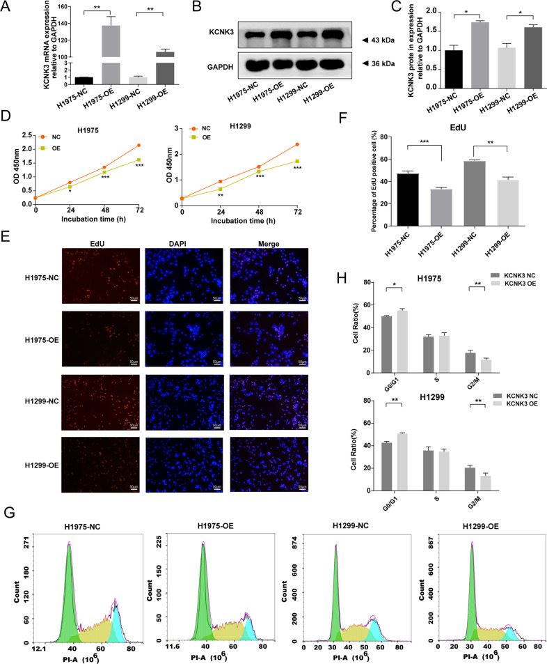
Fig. 2. Upregulation of KCNK3 expression inhibited the proliferation of LUAD cell lines.
A qRT-PCR analysis of KCNK3 expression was conducted after KCNK3 overexpression lentivirus transfection in H1975 cells and H1299 cells. B, C Relative KCNK3 protein expression was measured by western blot after transfection of KCNK3 overexpression lentivirus in H1975 cells and H1299 cells, with results normalized relative to the expression of GAPDH. D The effect of KCNK3 on LUAD cell viabilities was detected by CCK-8 assay and expressed as OD values. E, F EdU-594 staining assay was performed to evaluate the proliferation of KCNK3-overexpressed LUAD cells. The positive ratio was quantified by the counts of EdU-positive cells (red) and total counts of DAPI cells (blue), scale bar 50 µm. G, H The cell cycle of KCNK3 overexpression LUAD cells was analyzed using flow cytometry. (NC, negative control; OE, overexpression. Data are presented as the mean ± SD of three independent experiments. (*P < 0.05, **P < 0.01, ***P < 0.001).

