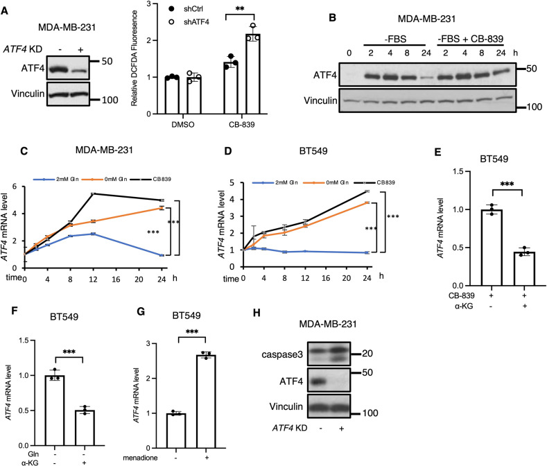
Fig. 1. ATF4 gene expression is the target of oxidative stress signaling in breast cancer.
A ROS detection assay in control or ATF4 KD MDA-MB-231 cells ± CB-839 (5 μM) for 24 h. Data shown are representative of two independent experiments and expressed as means ± SD for triplicate measurements. B Western blot analysis of whole cell lysates (WCL) from MDA-MB-231 cells ± CB-839 (1 μM) at indicated times. Blots are representative of two independent experiments. C, D RT-qPCR quantification of ATF4 mRNA in MDA-MB-231 and BT549 cells ± CB-839 (1 μM) or ± glutamine (2 mM) at the indicated times. Data shown are representative of two independent experiments and expressed as means ± SD for triplicate measurements. E, F RT-qPCR quantification of ATF4 mRNA in BT549 cells treated with CB-839 (1 μM) or without glutamine ± DM-αKG (2 mM) for 24 h. Data shown are representative of two independent experiments and expressed as means ± SD for triplicate measurements. G RT-qPCR quantification of ATF4 mRNA in BT549 cells treated with or without menadione (2 μM) for 24 h. Data shown are representative of three independent experiments and expressed as means ± SD for triplicate measurements. H Western blot analysis of WCL from MDA-MB-231 cells cultured in glutamine free condition for 48 h. Blots are representative of two independent experiments.

