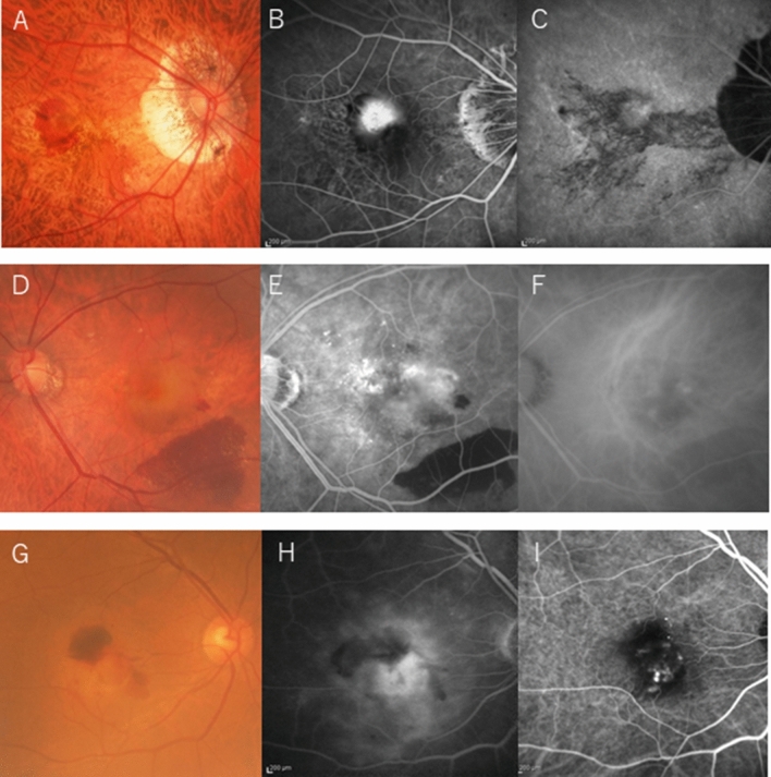
Figure 1.
Representative cases of the three groups. (A–C) The mCNV group has more severe chorioretinal atrophy than diffuse choroidal atrophy (A), fluorescein angiography (FA) shows classic CNV (B), and ICGA shows LCs (C). (D–F) The high-myopia CNV group shows no chorioretinal atrophy (MM less than category 2) (D), FA shows occult CNV and blockage due to subretinal hemorrhage (E), and ICGA shows polyp lesions (F). (G–I) The AMD group has an AL of less than 26.5 mm, no chorioretinal atrophy on fundus photography (G), occult CNV on FA (H), and polyp lesions on ICGA (I).

