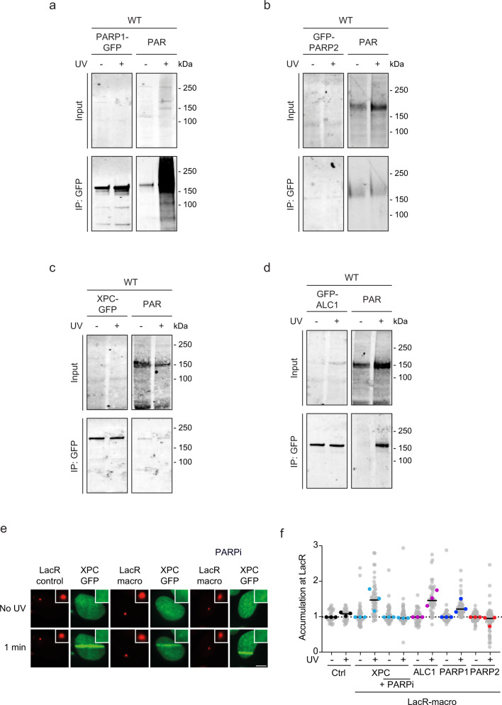
Fig. 5. PARP1 and ALC1 are PARylated in response to UV.
a–d Immunoprecipitation of a PARP1-GFP, b GFP-PARP2, c XPC-GFP, d GFP-ALC1 under high-salt conditions in the presence and absence of UV-C (20 J/m2, 15 min) stained for PAR (Millipore; MABE1016) or GFP. Three independent replicates of each IP experiment were performed obtaining similar results. e Representative images and f quantification of GFP-tagged XPC, ALC1, PARP1, or PARP2 recruitment to the LacO array upon tethering to the indicated mCherry-LacR-macrodomain. Pictures were taken before and 1 min after UV-C micro-irradiation. 24–45 nuclei were analyzed in three independent biological replicates (n = 3). Additional representative images are found in Fig. S3a. The scale bar in e is 5 µm.

