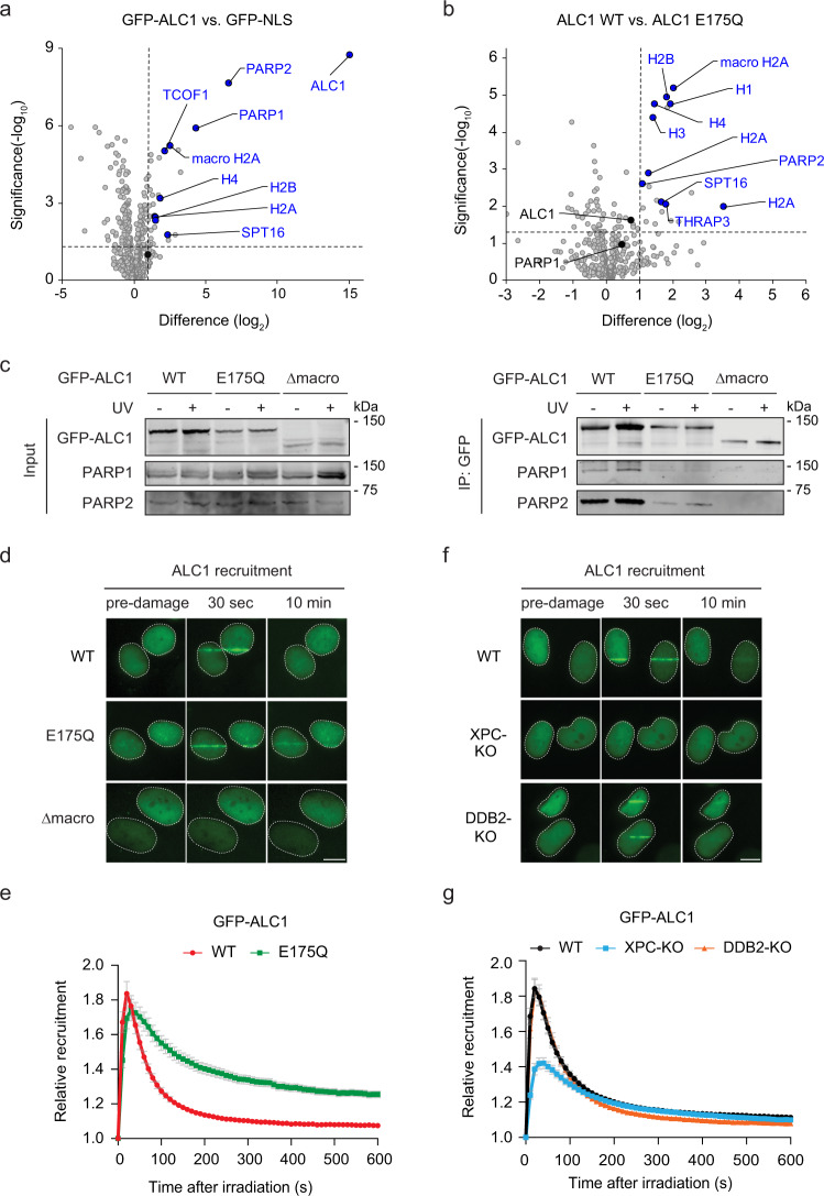
Fig. 7. The chromatin remodeler ALC1 is recruited to UV lesions by XPC.
a Volcano plot displaying the interactomes of GFP-ALC1 over GFP-NS after GFP-pull-down from U2OS (FRT) ALC1-KO cells and analysis by label-free proteomics. b Differential interactome of GFP-ALC1 WT vs. the catalytic-deficient GFP-ALC1 E175Q mutant. a, b The dashed lines indicate a twofold enrichment on the x axis (log2 of 1) and a significance of 0.05 (−log10 P value of 1.3; two-sided t test) on the y axis. c Co-IP of GFP-ALC1 WT, GFP-ALC1 E175Q, and a PAR-binding-deficient GFP-ALC1 Δmacrodomain in the presence and absence of UV-C (20 J/m2, 1 h). Three independent replicates of each IP experiment were performed obtaining similar results. d Representative images and e recruitment kinetics of GFP-ALC1 WT, GFP-ALC1 E175Q, GFP-ALC1 Δmacrodomain at sites of local UV-C laser irradiation in U2OS (FRT) ALC1-KO cells. 77–82 cells were analyzed in three independent experiments. f Representative images and g recruitment kinetics of GFP-ALC1 at sites of local UV-C laser irradiation in U2OS (FRT) WT, XPC-KO, and DDB2-KO cells. 107–135 cells were analyzed in n = 4 (WT), n = 5 (XPC-KO) or n = 6 (DDB2-KO) independent experiments. The data are shown as mean + SEM normalized to pre-damage GFP intensity at micro-irradiation sites. The scale bar in d, f is 5 µm.

