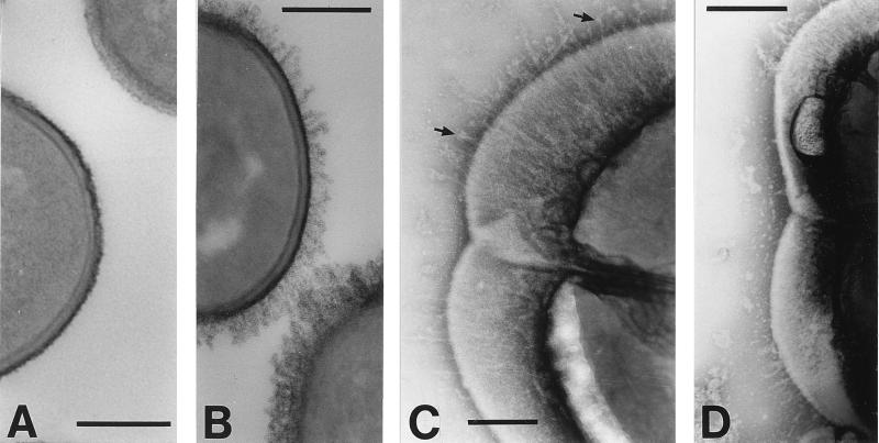
FIG. 5.
Electron microscopy of E. faecalis JH2-2 cells carrying pAM401 or pAMCshA. (A and B) Thin-section micrographs stained with RR and osmium tetroxide; (C and D) negatively stained cells. (A) E. faecalis OB513(pAM401) control cells are surrounded by a compact and densely stained layer covering a less-densely stained cell wall layer. (B) E. faecalis OB516(pAMCshA) cells expressing CshA polypeptide show a densely stained fibrillar fringe. (C) E. faecalis OB516 cells show peritrichous fibrils 70.3 ± 9.1 nm long that are more sparsely located in the region of the septum, and some fibrils have globular ends (arrows). (D) E. faecalis OB516 cells showing the absence of fibrils from the septal region and their expression restricted to the older ends of the cells. Bars, 200 nm.

