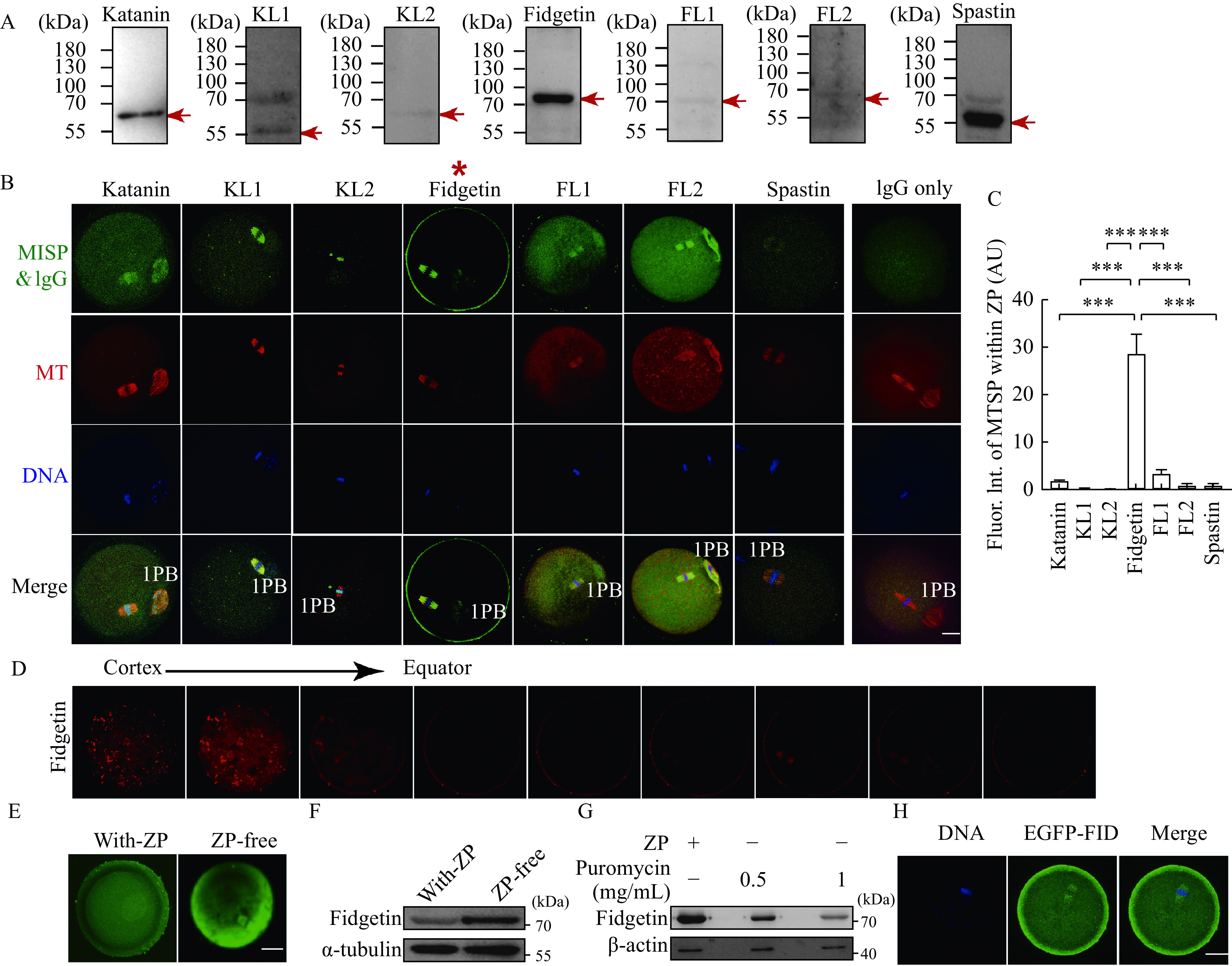
Figure 1.

Mouse fidgetin has a unique ZP-dominant localization among all MTSPs within oocytes.
A: The specificity of antibodies targeting the seven microtubule-severing proteins (i.e., katanin, katanin-like 1 [KL1], KL2, fidgetin, fidgetin-like 1 [FL1], FL2, and spastin) was identified by Western blotting. The red arrow indicates the band with molecular weight of each MTSP. B and C: The subcellular localization of each MTSP within mouse oocytes was examined by immunofluorescence (IF) staining, and MTSP fluorescence intensity within the zona-pellucida (ZP) was quantified. MTSPs in green, microtubule (MT) in red, and DNA in blue. Data are shown as mean±SEM.N=3, n=5 (oocytes) for C. Statistical analysis was performed using one-way ANOVA. ***P<0.001. 1PB: first polar body; Fluor: fluorescence; Int: intensity; AU: arbitrary unit. D: Z-series of fidgetin fluorescent image from ZP to the equator of the oocytes was aligned to show the enrichment of fidgetin within the ZP. E and F: IF staining and Western blotting were performed to show the change of fidgetin protein level when the ZP was removed by an acidic (pH 2.5) M2 medium. G: Western blotting assays were performed to show the change of fidgetin protein level when protein translation was inhibited by puromycin at the indicated concentrations for 1 hour before ZP removal. H:In vitro transcribed EGFP-fidgetin mRNA was injected into oocytes, and the localization of exogenous EGFP-fidgetin (EGFP-FID) was detected using an anti-EGFP antibody. Scale bar: 20 μm. α-tubulin or β-actin was used as the loading control.
