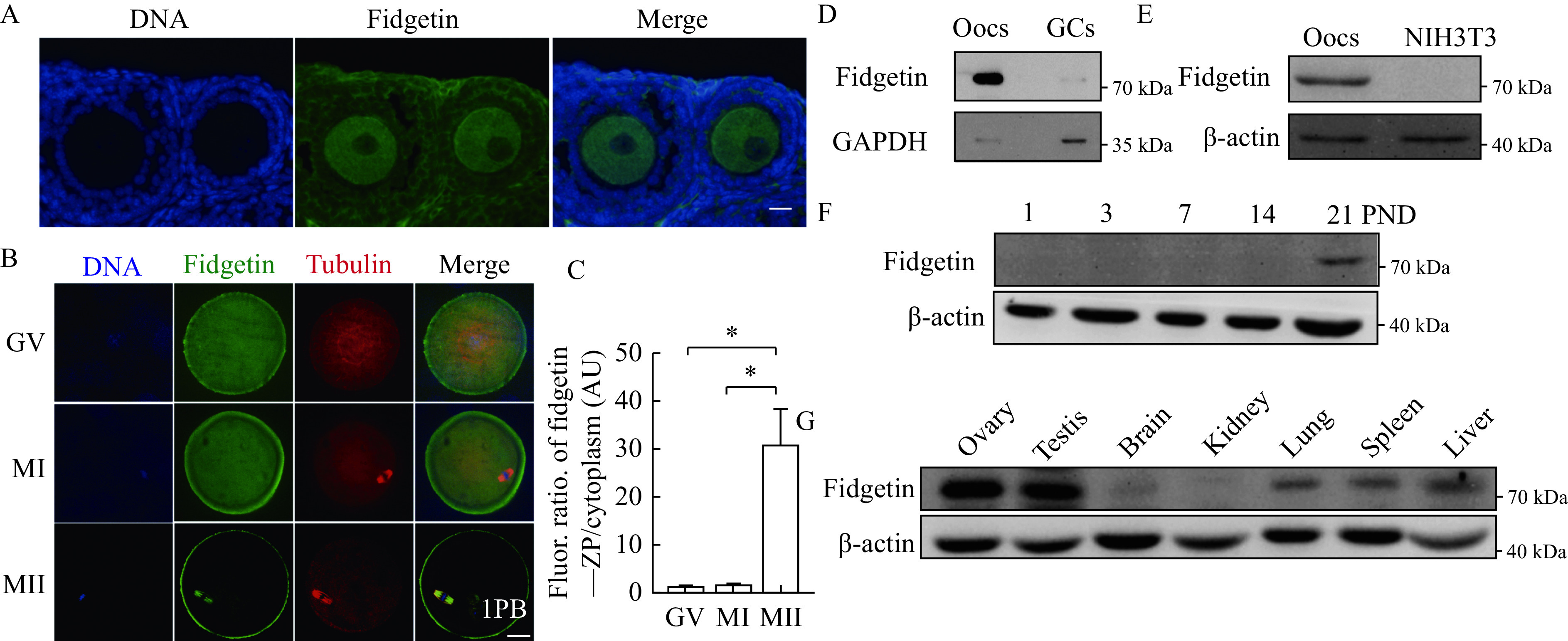
Figure 2.

Mouse fidgetin is enriched within ovaries and oocytes.
A: Fidgetin immunostaining within mouse ovaries was performed to show the expression of fidgetin within oocytes in antral follicles. DNA in blue and fidgetin in green. B and C: Immunostaining and quantification was performed to show the change of fidgetin during in vitro maturation from germinal vesicle (GV) to metaphase Ⅰ (MⅠ) and metaphase Ⅱ (MⅡ). DNA in blue, fidgetin in green, and tubulin in red. 1PB: first polar body. Fluor: fluorescence; ZP: zona pellucida; AU: arbitrary unit. Data are shown as mean±SEM. N=3 for C. Statistical analysis was performed using one-way ANOVA. *P<0.05. D: Western blotting was performed to show the expression of fidgetin within oocytes (Oocs) and granulosa cells (GCs). E: Western blotting was performed to show the expression of fidgetin within Oocs and somatic cells (NIH3T3). F: Western blotting was performed to show the fidgetin levels within mouse ovaries with different postnatal days (PNDs). G: Western blotting was performed to show the expression of fidgetin within mouse ovaries, testis and other tissues. Scale bar: 20 μm. GAPDH or β-actin was used as the loading control.
