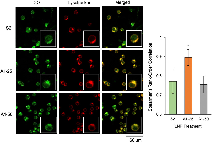
Fig. 5.
Endosomal uptake and colocalization of mRNA LNPs with varying 7α-hydroxycholesterol substitutions with endosomes in Jurkats. Confocal microscopy images of Jurkat cells treated with DiO-labeled LNPs at 60 ng mRNA/ 60,000 cells and stained with Lysotracker. Images were merged, background was subtracted, and Spearman's rank-order correlation was used to quantify association between LNPs and acidic organelles in cells. Single cell insets have been included for each image to provide visual reference, and insets are 2× magnification of the original image. Colocalization statistics (i.e., Spearman's rank-order correlation) were obtained from 5 fields of view (at least 90 cells in total) of each treatment group. Error bars denote standard deviation. An ANOVA was performed to determine if group means differed significantly. *: p < 0.05 in student t-test with Bonferroni p-value correction between colocalization statistics from S2 and either A1–25 or A1–50 LNPs.

