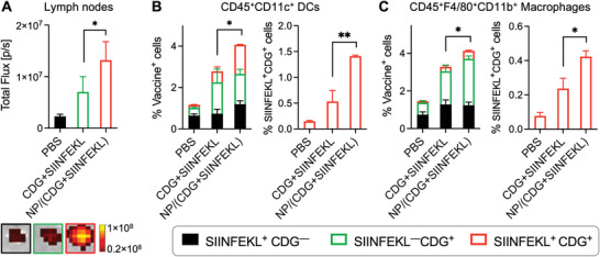
Figure 4.

Nanovaccines (NVs) improved the codelivery of DY547‐CDG and SIINFEK(FITC)L to draining lymph nodes (LNs) and intranodal antigen presenting cells (APCs). A) Signal quantification (left) and representative photos (right) of draining inguinal LNs 18 h after s.c. administration of NVs or a soluble mixture of SIINFEKL and CDG at tail base in C57BL/6 mice (n = 3). B,C) Flow cytometry data quantification showing the NV codelivery of CDG and SIINFEKL into LN‐residing dendritic cells (DCs) (B) and macrophages (C), two primary intranodal APC subsets. Data: mean ± SEM (n = 3); *p < 0.05, **p < 0.01, ***p < 0.001, and ****p < 0.0001 (Student's t‐test).
