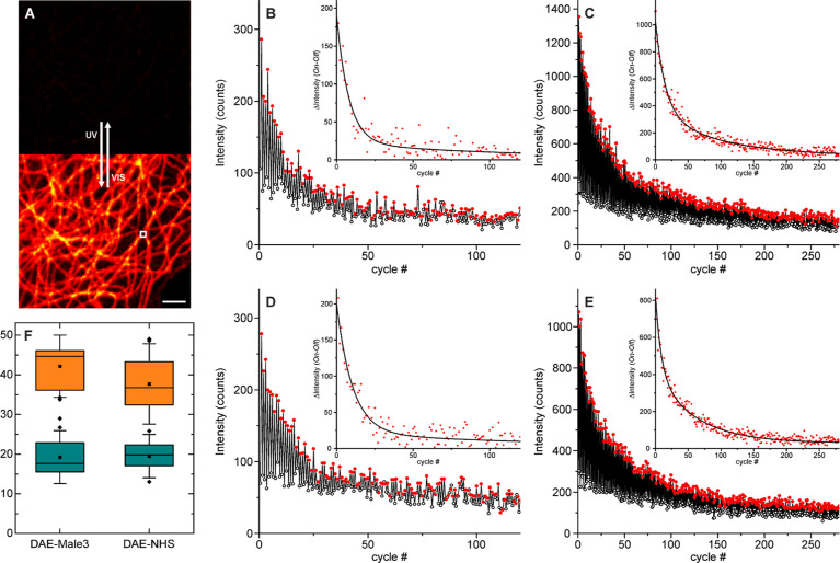
Figure 5.
Photoswitching fatigue resistance observed on a confocal microscope for the labeled secondary antibodies on microtubules on fixed Cos7 cells. (A) Confocal image without and with activation (355 nm) after a switching experiment was performed in the indicated area (white square). Switching on and off on a sample labeled with DAE-Male3 mounted in PBS without (B) and with (C) CB7 (2 mM) and on a sample labeled with DAE-NHS without (D) and with (E) CB7 (2 mM). The insets show the differences between two successive substeps (symbols) along with a biexponential fit (lines). (F) Boxplots of the mean (amplitude averaged) characteristic switching time (20 measurements on different positions for each case). Scale bar on A: 2 μm.

