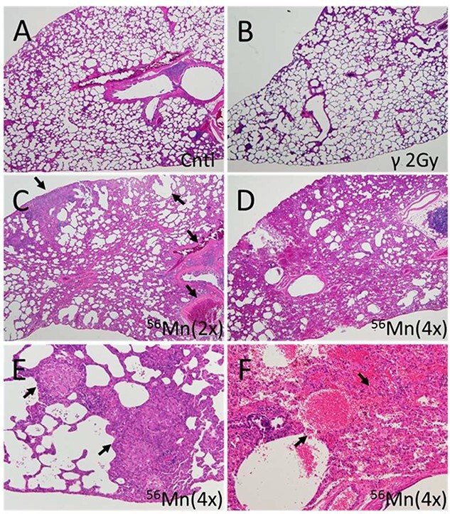
Fig. 4.

Representative fields of lung tissue histology from 56Mn-exposed, 2.0-Gy γ-exposed and nonirradiated control rats six months post-irradiation. (A) Lung of a control rat showing no pathological changes; (B) lung of a 2.0-Gy γ-radiation exposed rat showing no pathological changes; (C) lung of a 56Mn(2×) exposed rat showing widespread emphysema, atelectasis, hemorrhage and thrombosis (arrows); (D) lung of a 56Mn(4×) exposed rat showing more extensive damage and pneumonitis due to severe inflammation, inflammatory cell infiltration and intra-alveolar hemorrhage; (E) lung of a 56Mn (4×) exposed rat showing granuloma surrounded by emphysema in a lung lobe (arrows); and (F) lung of a 56Mn (4×) exposed rat showing severe hemorrhage and inflammatory cell infiltration (arrows) and this caption is a higher magnification of caption (D). H&E stain, 4× (A–D) and 20× (E, F) objective magnification (From [6]/CC BY 4.0).
