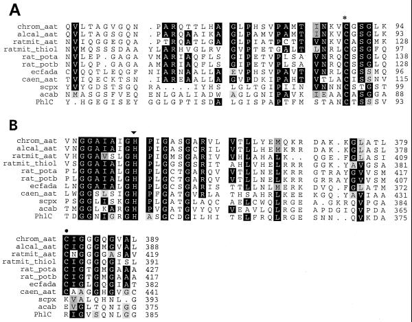
FIG. 2.
Important residues conserved in PhlC. (A) Alignment of cysteine 88 (∗) of PhlC with the catalytic active site cysteine of thiolases. (B) Alignment of histidine 349 (▾) and neighboring glycine residues of PhlC. Cysteine 378 (●), a key active site residue in type II thiolases, is absent from PhlC, SCPx, and AcaB. In both panels, the compared proteins are chrom_aat, Chromatium vinosum acetoacetyl-CoA thiolase (accession. no. L01112); alcal_aat, Alcaligenes eutrophus acetoacetyl-CoA thiolase (accession no. J04987); ratmit_aat, rat mitochondrial acetoacetyl-CoA thiolase (accession no. D13921); ratmit_thiol, rat mitochondrial oxoacyl-CoA thiolase (accession no. X05341); rat_pota, rat peroxisomal oxoacyl-CoA thiolase A (accession no. M32801); rat_potb, rat peroxisomal oxoacyl-CoA thiolase B (accession no. J02749); ecfada, E. coli 3-ketoacyl-CoA thiolase (accession no. P21151); caen_aat, Caenorhabditis elegans acetyl-CoA acetyltransferase (accession no. M77697); scpx, mouse sterol carrier protein 2 precursor (accession no. P32020); and acab, P. furiosus acetyl-CoA synthase. Numbers to the right of the alignment are amino acid residue numbers.

