
FIGURE 1.
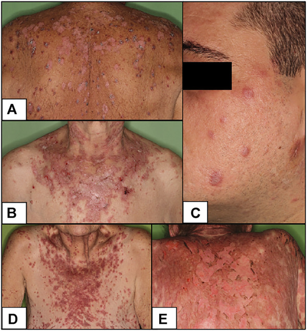
Clinical pictures of our patients (A) DI-SCLE (Case 1) (B) I-SCLE (Case 3) (C) DLE (Case 6) (D,E) TEN-like (ACLE) lupus early and late (Case 9 and 10) Clinical presentations of DI-SCLE and SCLE cases reveal similarities: scaling, erythematous plaques in sun-exposed body parts. Clinical picture of DLE shows the deeply infiltrated, discoid plaques on the face. The two pictures of the TEN-like lupus case present the progression of the clinical picture (SCLE, subacute cutaneous lupus erythematosus; DI-SCLE, PD-1 inhibitor-induced SCLE; I-SCLE, idiopathic SCLE; DLE, discoid lupus erythematosus; TEN, toxic epidermal necrolysis).