
FIGURE 2.
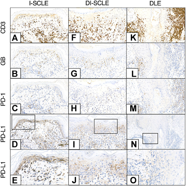
CD3, GB, PD-1, PD-L1 immunohistochemistry of I-SCLE, DI-SCLE and DLE (A–E) I-SCLE (F–J) DI-SCLE (K–O) DLE [Magnifications: ×20, except: (N), ×10; (E,J,O): ×40] The immunohistochemical pictures of DI-SCLE and I-SCLE do not show many differences, however dermal GB cell number is higher in I-SCLE. In DLE a more massive and perifollicular dermal infiltrate can be observed compared to SCLEs. KC PD-L1 expression is elevated in all cases, however a high increase can be seen in SCLEs (regardless of origin) while only slight increase in DLE (SCLE, subacute cutaneous lupus erythematosus; DI-SCLE, PD-1 inhibitor-induced SCLE; I-SCLE, idiopathic SCLE; DLE, discoid lupus erythematosus; GB, granzyme B; KC, keratinocyte; PD-1, programmed cell death 1; PD-L1, programmed cell death ligand 1).