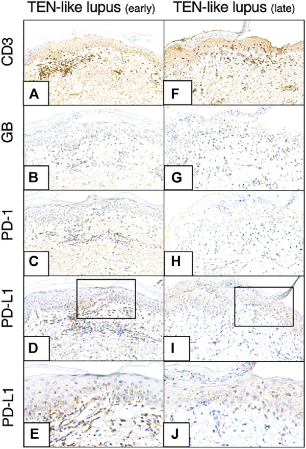
FIGURE 4.

CD3, GB, PD-1, PD-L1 immunohistochemistry of earlier and later TEN-like lupus samples (A–-E) TEN-like lupus, early sample (F–J) TEN-like lupus, late sample [Magnifications: ×20, except: (E,J): ×40] As the clinical picture progressed activated T-cell numbers (GB+ T-cell numbers) and KC PD-L1 expression increased (TEN, toxic epidermal necrolysis; GB, granzyme B; KC, keratinocyte; PD-1, programmed cell death 1; PD-L1, programmed cell death ligand 1).
