. 2022 Mar 3;37(2):195–204. doi: 10.46497/ArchRheumatol.2022.8252

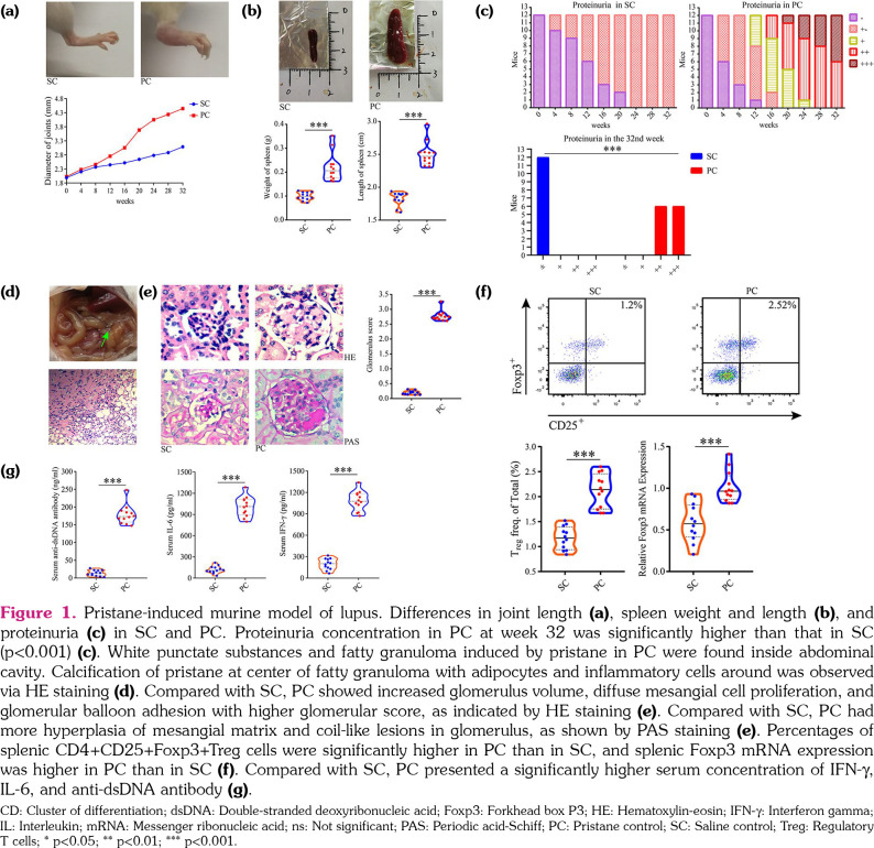
Figure 1. Pristane-induced murine model of lupus. Differences in joint length (a), spleen weight and length (b), and proteinuria (c) in SC and PC. Proteinuria concentration in PC at week 32 was significantly higher than that in SC (p<0.001) (c). White punctate substances and fatty granuloma induced by pristane in PC were found inside abdominal cavity. Calcification of pristane at center of fatty granuloma with adipocytes and inflammatory cells around was observed via HE staining (d). Compared with SC, PC showed increased glomerulus volume, diffuse mesangial cell proliferation, and glomerular balloon adhesion with higher glomerular score, as indicated by HE staining (e). Compared with SC, PC had more hyperplasia of mesangial matrix and coil-like lesions in glomerulus, as shown by PAS staining (e). Percentages of splenic CD4+CD25+Foxp3+Treg cells were significantly higher in PC than in SC, and splenic Foxp3 mRNA expression was higher in PC than in SC (f). Compared with SC, PC presented a significantly higher serum concentration of IFN-g, IL-6, and anti-dsDNA antibody (g). CD: Cluster of differentiation; dsDNA: Double-stranded deoxyribonucleic acid; Foxp3: Forkhead box P3; HE: Hematoxylin-eosin; IFN-g: Interferon gamma; IL: Interleukin; mRNA: Messenger ribonucleic acid; ns: Not significant; PAS: Periodic acid-Schiff; PC: Pristane control; SC: Saline control; Treg: Regulatory T cells; * p<0.05; ** p<0.01; *** p<0.001.

Figure 1