
Fig. 7. dACE2 is non-functional for binding SARS-CoV-2 spike protein RBD and as a carboxypeptidase.
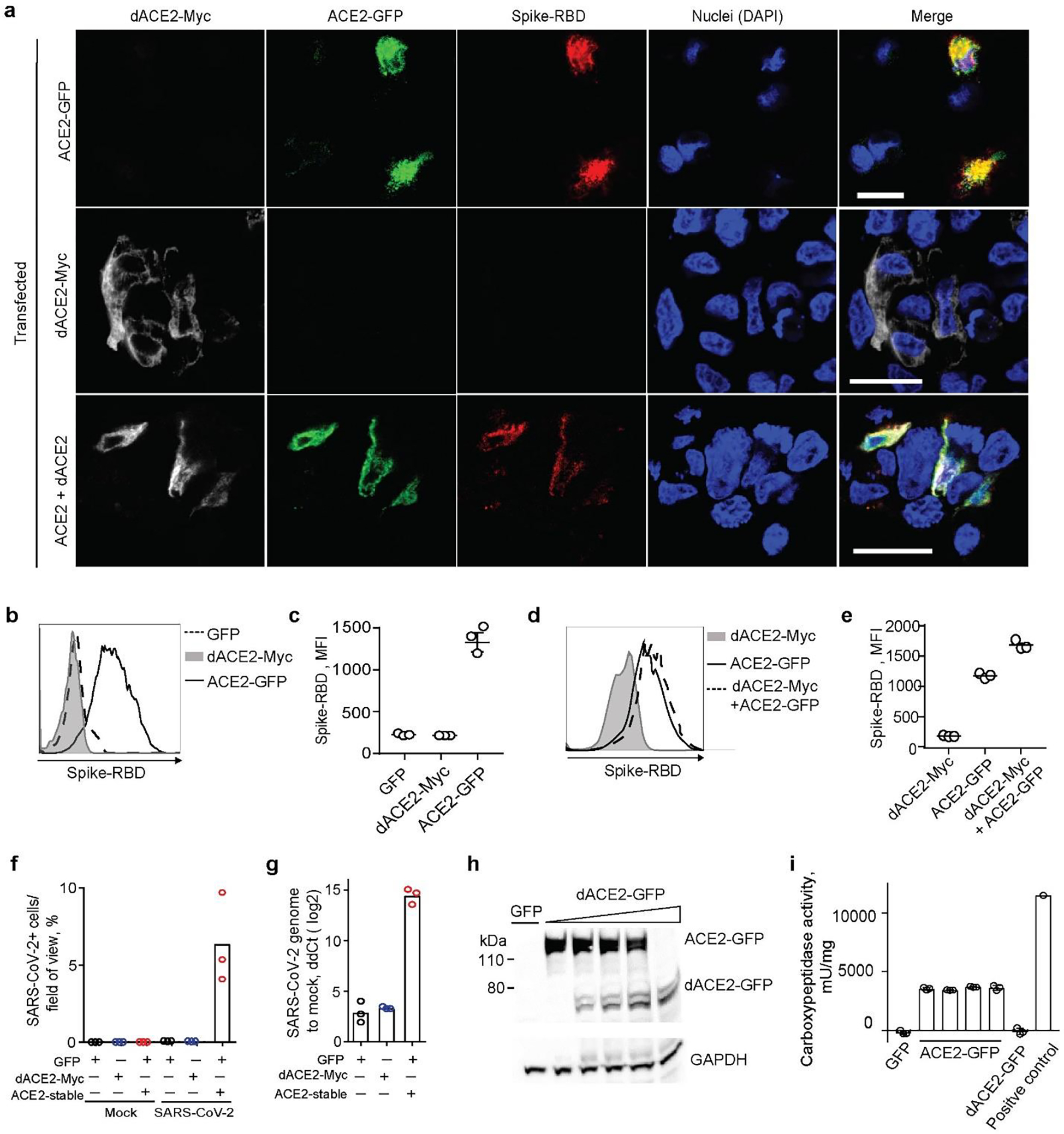
a, Representative confocal images of T24 cells transiently overexpressing dACE2-Myc (white), ACE2-GFP (green) and treated with receptor-binding domain (RBD) of SARS-CoV-2 spike protein (red), nuclei (DAPI)-blue; bars=20μM. b-d) Representative flow cytometry histogram b, and mean fluorescence intensity (MFI) values from 3 biological replicates c, of spike-RBD binding to the surface of ACE2-GFP but not dACE2-Myc expressing T24 cells. Gating for cells expressing dACE2-Myc, ACE2-GFP, or both proteins, is shown in Extended Data Fig. 9a. e, plot depicting MFI of spike protein-RBD binding. The results are based on 3 biological replicates, shown one of two independent experiments. f, SARS-CoV-2 infectivity rates (%) in a lung cancer cell line A549 transfected with GFP, or dACE2-Myc, or stably expressing ACE2 (ACE2-stable) and transfected with GFP. g, SARS-CoV-2 viral load as ddCt values compared to mock, corresponding to plot f. Additional details are provided in Extended Data Fig. 10. h, A representative Western blot with an anti-ACE2 antibody that detects both recombinant ACE2-GFP and dACE2-GFP overexpressed in T24 cells. The amount of the ACE2-GFP lysate was kept constant, while the amount of dACE2-GFP cell lysate was increased and the difference in the lysate volume was compensated by the empty GFP vector. i, Results of carboxypeptidase assays using variable amounts lysates of cells (as described in plot H, showing that the activity of ACE2 is not affected by increasing amounts of dACE2. The results are based on 3 biological replicates and presented with means and SD. WB is showing the results of one representative replicate.