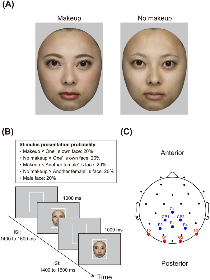
Fig 3. Stimuli and procedures in Experiment 2.
Panel A depicts an example of facial images. The individual in this figure has given written informed consent to publish her facial images. Panel B illustrates a schematic representation of the gender classification task. Panel C shows the EEG sensor layout and ROIs. The electrodes included in the occipitotemporal ROI are marked in red, and the blue circles indicate electrodes located in the centroparietal ROI. EEG, electroencephalogram; ROI, region of interest.

