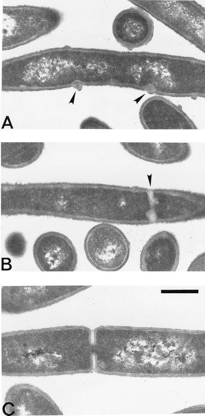
FIG. 6.
Electron micrograph showing cross-sections of ponA mutant and wild-type cells grown at 37°C in 2× YT medium. (A and B) ponA mutant cells; (C) wild-type cell. The arrowheads indicate aberrant septa or cell wall clusters in ponA mutant cells. Note that the cell wall clusters shown in panel A are at positions overlapping the DNA. Bar, 0.5 μm.

