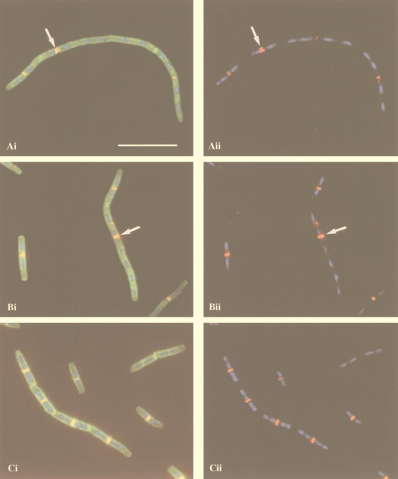
FIG. 7.
Immunolocalization of FtsZ in ponA mutant (A and B) and wild-type (C) cells grown in 2× YT medium at 37°C. (Ai, Bi, and Ci) Overlay of FtsZ localization (red and yellow), DAPI staining of nucleoids (blue), and staining of cell walls and septa with FITC-conjugated WGA (green); (Aii, Bii, and Cii) overlay of FtsZ localization (red) and DAPI staining (blue). The arrows in panels Ai and Aii indicate an aberrant FtsZ ring, and those in panels Bi and Bii indicate an FtsZ ring at an inappropriate location. Bar, 10 μm.

