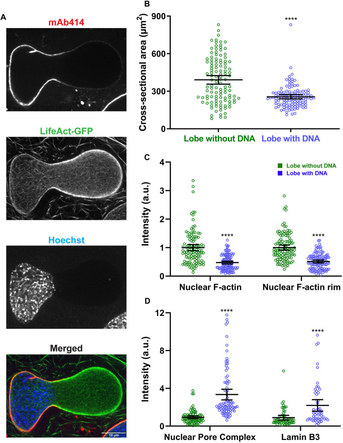
Fig. 2.
The nuclear envelope is structurally heterogeneous in bilobed nuclei. (A) After 90 min of nuclear assembly in F-actin-intact extracts, nuclei were stained with mAb414 (to label NPCs), LifeAct–GFP (F-actin) and Hoechst 33342 (DNA) and imaged by confocal microscopy. In some cases, F-actin was labeled with Alexa Fluor 488 phalloidin. A bilobed nucleus is shown (representative of four experiments). (B) Nuclear cross-sectional area was quantified based on F-actin staining for the lobes with and without Hoechst 33342-stained DNA (n=113 nuclei, five independent experiments) using widefield microscopy. (C) Total nuclear F-actin and F-actin localized to the nuclear rim were quantified (n=113 nuclei, five independent experiments) using widefield microscopy, in each case normalized to the lobe without Hoechst 33342-stained DNA. (D) NPC intensity in the two lobes was quantified based on mAb414 staining (n=85 nuclei, four independent experiments) using widefield microscopy. To quantify Lamin B3 intensity in the two lobes, extracts were supplemented with GFP–Lamin B3 and imaging performed by confocal microscopy (n=54 nuclei). In both cases, intensity values were normalized to the lobe without Hoechst 33342-stained DNA. Mean values and 95% c.i. error bars are shown. Nonparametric Mann–Whitney tests were performed, showing statistical significance relative to the lobe without Hoechst 33342-stained DNA. a.u., arbitrary units. ****P≤0.0001.

