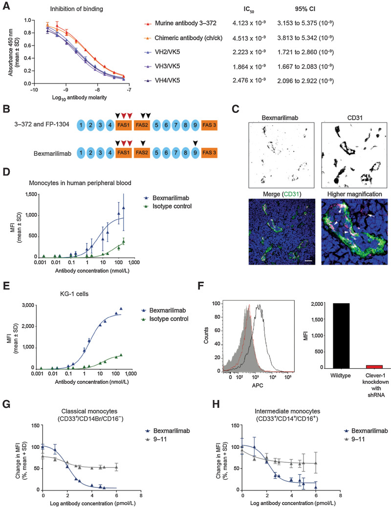
Figure 1.
Characterization of bexmarilimab binding in vitro. A, Binding of biotinylated parental anti–Clever-1 antibody 3–372 to human Clever-1 in the presence of nonbiotinylated 3–372, chimeric anti–Clever-1 antibody and three composite antibodies (VH2/VK5, VH3/VK5, VH4/VK5) as determined by a competitive ELISA. IC50 values were calculated by three-parameter nonlinear curve fitting with 95% confidence intervals (CI). B, Bexmarilimab-recognized region of Clever-1. The orange boxes and blue closed circles indicate fasciclin domains and EGF-like domains, respectively. The arrowheads indicate relative positions of identified binding motifs, the core epitopes are shown in red. C, Bexmarilimab immunoreactivity (magenta) in human lymph node with CD31 (green) and Hoechst (blue) co-staining. Scale bar 40 μm. Arrowheads point to Clever-1 expression on lymphatic endothelial cells and the arrow points to a single CD31-negative cell. Binding of bexmarilimab to human CD14+ cells (D) and human KG-1 acute myelogenous leukemia cells (E) as determined by flow cytometry. F, Binding and quantitation of bexmarilimab and the isotype control to KG-1 cells transfected with a Clever-1–targeting shRNA as determined by flow cytometry. The wild-type histogram is shown in black, the Clever-1 knockdown histogram in red, and the isotype control binding in solid gray. APC, allophycocyanin. Receptor occupancy of bexmarilimab in classical (G) and intermediate monocytes (H), shown as change in fluorescence intensity of CD14+ cells that bind to labeled bexmarilimab or mAb 9–11. Representative data are shown for 1 of 3 donors. MFI, mean fluorescence intensity.

