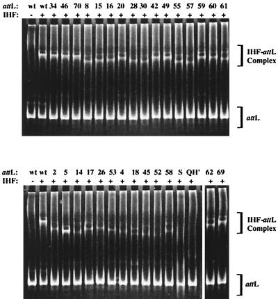
FIG. 2.
EMSA of attL with IHF. Each attL (200 nM) was incubated in the presence of IHF (60 nM) for 40 min at 25°C. Each reaction mixture was then resolved by polyacrylamide gel electrophoresis. The gels were stained in ethidium bromide and visualized under 302-nm light. Complexed DNA has a retarded migration pattern relative to free DNA. Numbers atop each lane indicate the attL. Lanes marked wt contain wild-type attL. Lane S contains the original attL population from which selection was performed. Lane QH′ contains attL with four base mutations in the 3′ binding domain (see the text).

