
Figure 4.
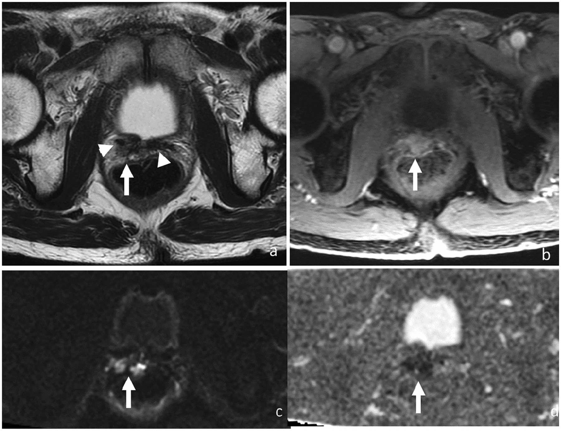
Axial T2-weighted (a), DCE (b), DWI (c) and ADC (d). 68-year-old man with rising PSA (5.78 ng/ml) 6 years after radical prostatectomy for Gleason score 4+4 prostate cancer. MRI shows a 2.2-cm T2 intermediate signal mass (arrow) in the right prostatectomy bed scar tissue demonstrating T2 low signal (arrowheads). Mass shows early enhancement and restricted diffusion. Patient was started on androgen deprivation therapy after which both this recurrent tumor on MRI and PSA levels decreased.