
Figure 5.
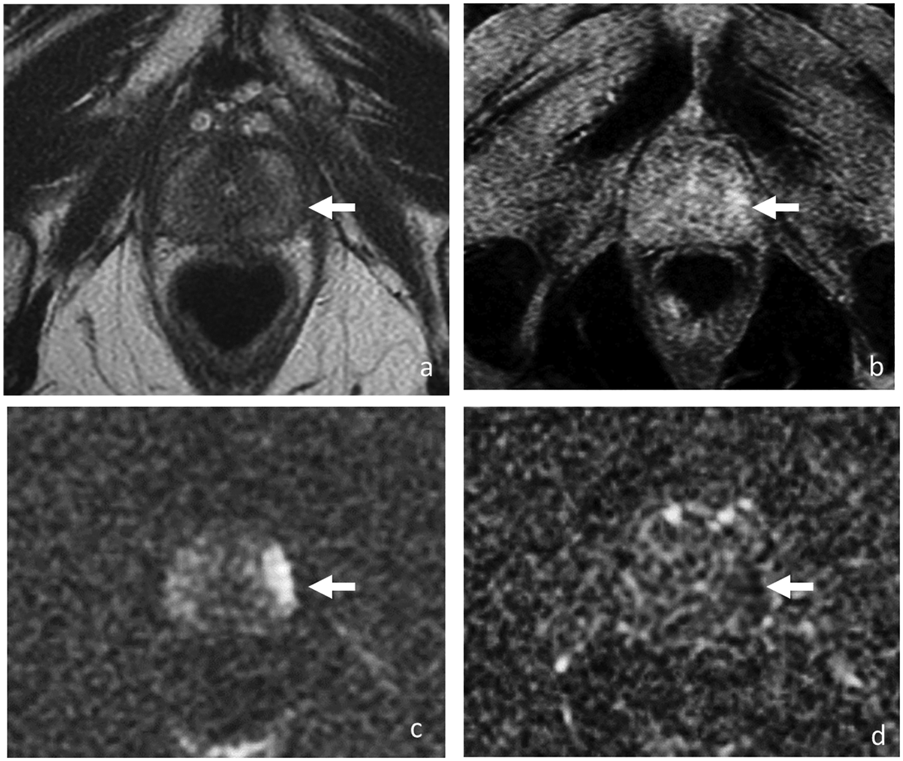
Axial T2-weighted image (a), DCE (b), DWI (c) and ADC (d). 64-year-old man with rising PSA biochemical recurrence (3.59 ng/ml) after external beam radiotherapy to a Gleason 3 + 3 prostate cancer 14 years ago. Diffuse low T2 signal throughout entire prostate and loss of zonal differentiation represent post-treatment changes, limiting detection of recurrent tumor. However, 1.6-cm focal lesion (arrow) in the left mid gland peripheral zone is demonstrated on early DCE images and diffusion-weighted images which was confirmed on biopsy as Gleason 4 + 4 cancer.