
Figure 6.
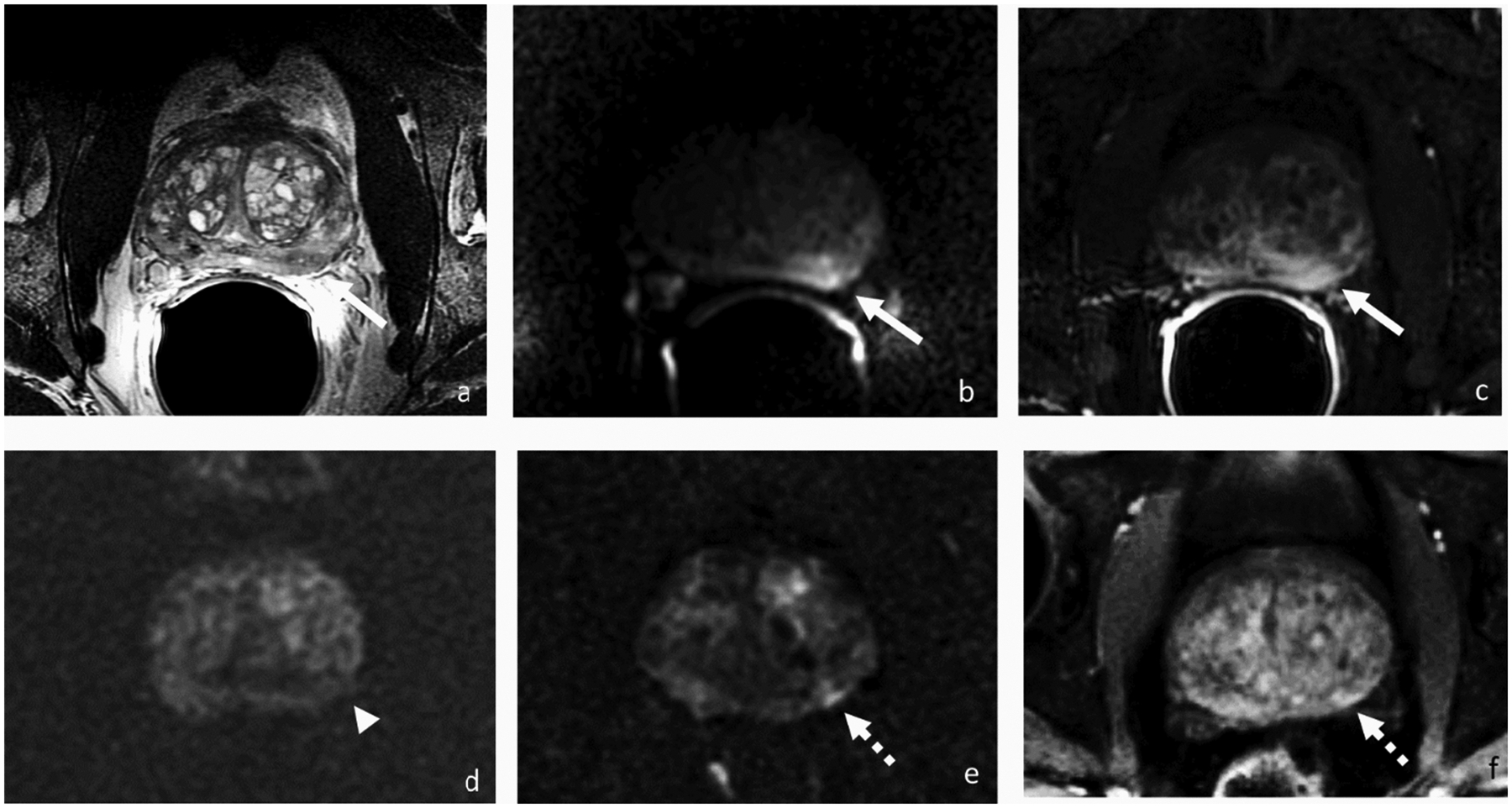
Axial T2-weighted (a), DWI (b) and DCE-MRI (c) before, DWI (d) 3 months after, and DWI (e) and DCE-MRI (f) 2 years after focal therapy, respectively. 63-year-old patient with PSA 5.8 ng/ml and a left mid gland peripheral zone lesion at presentation for which biopsy showed Gleason 3+4 prostate cancer. Three months after irreversible electroporation (IRE), there was no abnormal signal on DWI at the site of the treated tumor (arrowhead). Two years after IRE with PSA rose to 10.76 ng/ml, and MRI demonstrated a lesion (broken arrow) adjacent to the ablation site demonstrating early enhancement and restricted diffusion suspicious for recurrence that was confirmed by biopsy.