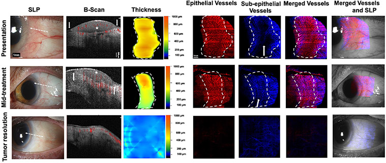
Fig. 6. A 69-year-old woman presenting with a gelatinous and papillomatous OSSN of the left eye (Case 7). SLP:
A gelatinous and papillomatous OSSN of the conjunctiva encroaching on the cornea temporally from 12 to 6 o’clock in the left eye with intrinsic vascularity and feeder vessels is seen at presentation. At mid-treatment, after 2 cycles of 5-FU, the gelatinous and papillomatous OSSN has dramatically decreased in size. At tumor resolution, the conjunctiva appears normal while some opacity (consistent with scar) remains in the cornea temporally. White dashed arrow shows cross-sectional cut of OCT. B-scan images: At presentation, thickened hyperreflective epithelium (asterisk) with abrupt transition (arrows) is seen. Blood flow (shown in red) within the tumor as well as in the subepithelium is noted. Some shadowing limits complete blood flow visualization under the tumor. At mid-treatment, the flow in the epithelium and subepithelium is still present. At tumor resolution, thin epithelium is noted and blood flow in subepithelium is normalized. Thickness map: Changes in the thickness of the tumor are shown at presentation, mid-treatment, and at tumor resolution. Dashed white lines indicate the tumor boundary. Enface angiograph of epithelial vessels: Dense BV in a “sea fan” pattern seen at presentation, decreasing at mid-treatment and disappearing at tumor resolution. Straight horizontal lines represent artifacts due to patient movement. Dashed white lines indicate the tumor boundary. Enface angiograph of the subepithelial vessels (200 μm): Dense BV at and adjacent to the tumor edge are seen at presentation. Complete visualization of BV immediately underneath the tumor is blocked due to the thick size of the tumor. At mid-treatment, BV adjacent to the tumor appear similar to presentation. In the contrary, BV immediately beneath the tumor are now more visible as the tumor is decreasing in thickness. With tumor resolution, BV adjacent to the tumor and immediately beneath the tumor disappeared. At resolution, subepithelial VAD levels below the tumor were comparable with subepithelial VAD levels in a similar location in the contralateral uninvolved eye. Dashed white lines indicate the tumor boundary. Merged vessels: The tumor BV (red) and subepithelial BV angiographies (blue) were merged at presentation, mid-treatment and at tumor resolution. Dashed white lines indicate the tumor boundary. Merged vessels and SLP: Overlay of the enface angiograph with grayscale slit-lamp at presentation, mid-treatment and at tumor resolution, showing gradual resolution of the epithelial vasculature and normalization of the subepithelial BV.OSSN; ocular surface squamous neoplasia, SLP; slit lamp photograph, OCT; optical coherence tomography, BV; blood vessels.

