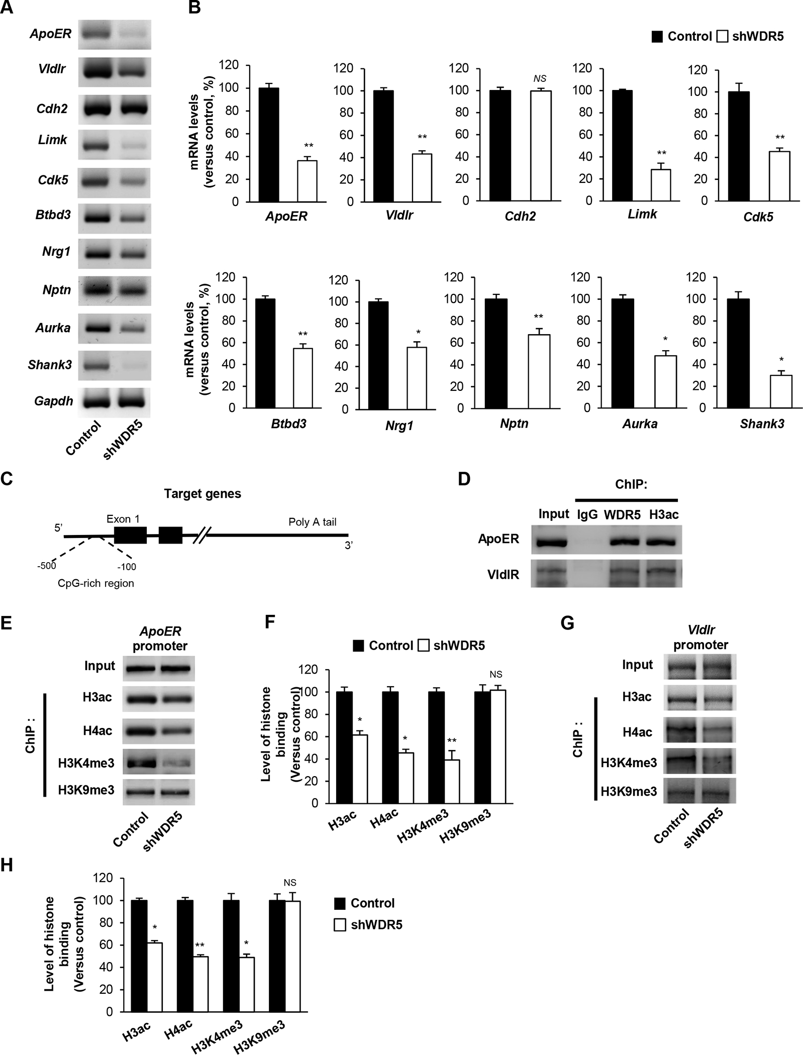
Figure 5. WDR5 regulates reelin signaling via regulating reelin signaling components expression.

(A) Knockdown of WDR5 decreases the transcript levels of Reelin signaling components and neurite development related genes. Cortical neurons from E15.5 mice were cultured and infected with a lentivirus encording control or shWDR5 at 5 DIV. Cellular lysates from the cultures at 14 DIV were subjected to RNA isolation and RT-PCR. (B) Quantification of transcript levels of (A). The relative levels of the genes were normalized to GAPDH expression. The band intensities were measured using Image J software. n=3 independent primary cultured cortical neurons using 3 mice. Statistical significance was determined by two-tailed Student’s t-test. *p < 0.05, **p < 0.01. (C) Illustration showing the CHIP assay of target gene promoter region. (D) WDR5 directly interacts with ApoER, VldlR and CDK5 gene promoter. ChIP assays were performed using primary cultured neurons. (E), (G) WDR5 suppression significantly decrease the level of acetylated Histone H3 at the target genes promoter. (F), (H) Quantification of (E), (G). The relative levels of the fold change were normalized to input fraction of Acetyl-histone H3 levels at the target genes promoter. n=3 independent primary cultured cortical neurons using 3 mice. Statistical significance was determined by two-tailed Student’s t-test. *p < 0.05, **p < 0.01.
