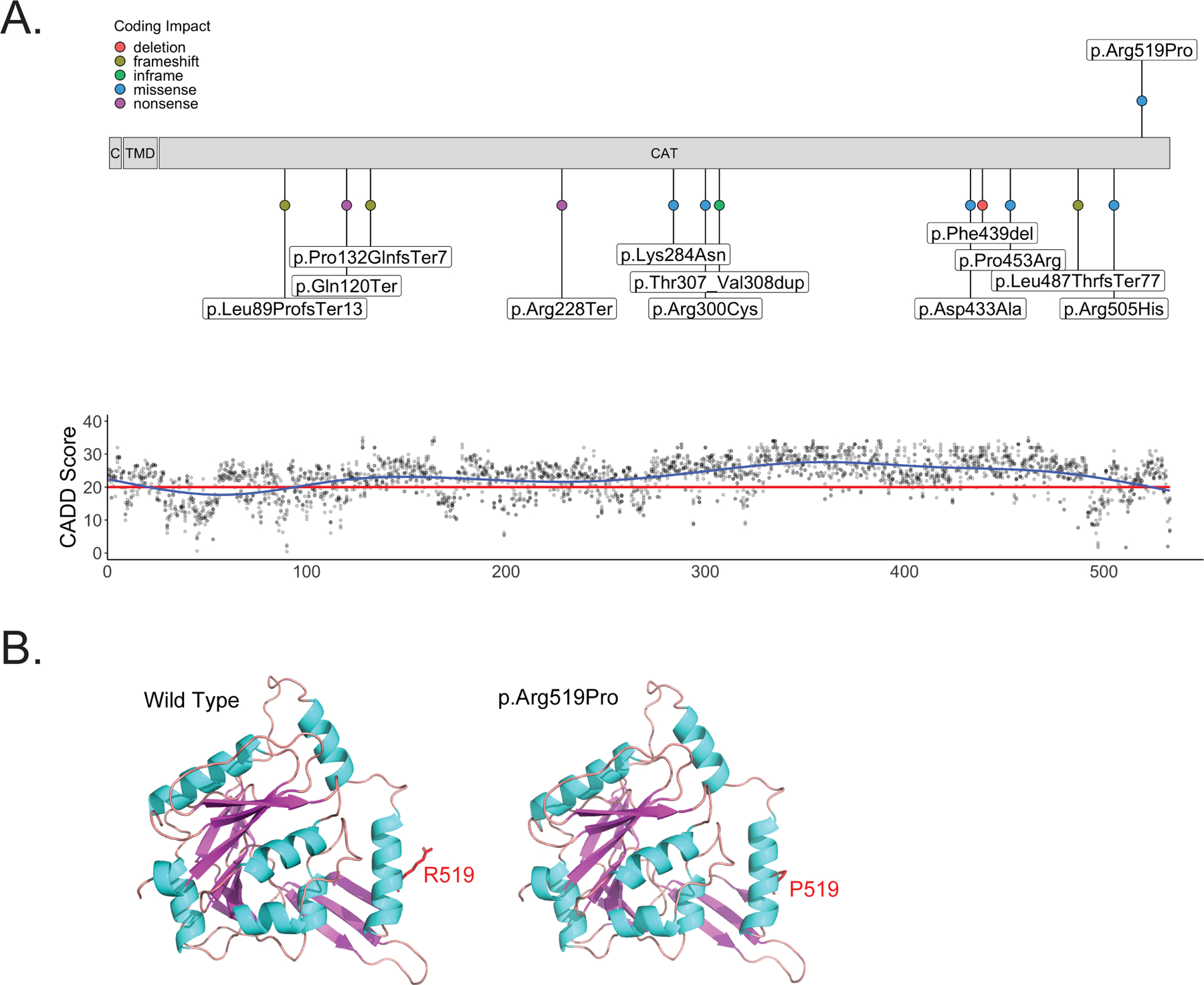
Figure 1.

B4GALNT1 protein structure with variant distribution.
A) Schematic of B4GALNT1 protein structure. The p.Arg519Pro missense variant discovered in the proband is shown above the schematic. All previously published exonic variants are shown below the schematic. The lower panel shows CADD PHRED v1.6 values for all possible missense variants in B4GALNT1 across the linear protein structure. The red line marks the recommended cut off of 20. B) 3D structural model of the B4GALNT1 catalytic domain (273–533) derived from the AlphaFold database (left) and a model of the p.Arg519Pro variant generated using Discovery Studio (right). The 3D structures are shown in a ribbon model and Arg519 (left) and Pro519 (right) are shown in stick model. The figure was prepared using PyMol. Since proline is established as a helix breaker, the arginine-to-proline mutation is predicted to destabilize the helix structure.
