FIGURE 3.

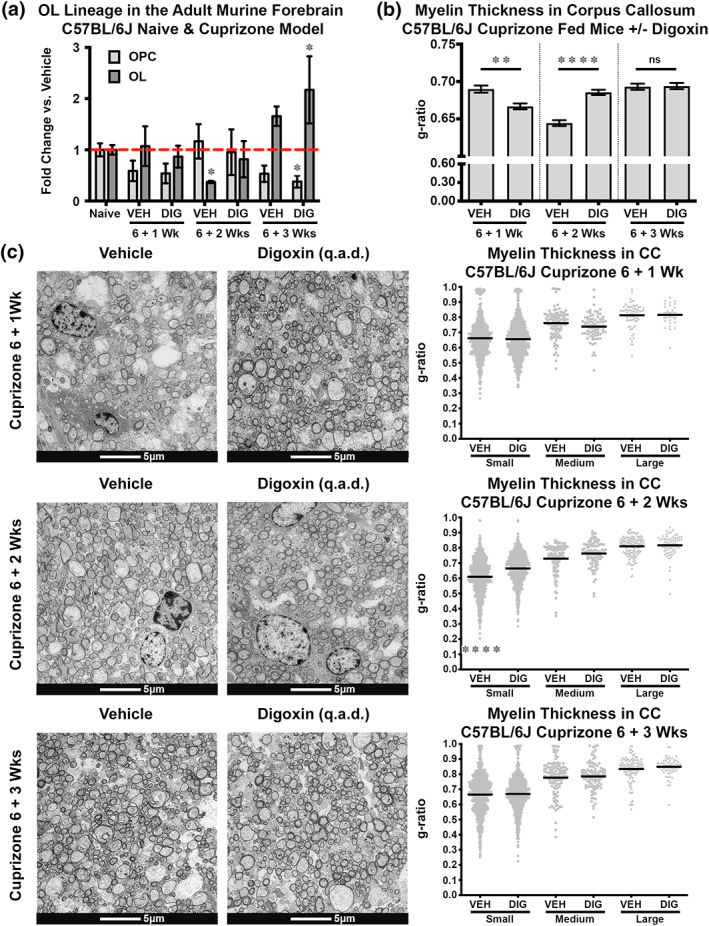
Digoxin treatment promoted earlier myelin regeneration in the brains of female C57BL/6J mice following cuprizone‐induced demyelination. 8‐week‐old female C57BL/6J mice were fed control or cuprizone chow for 6 weeks and then placed on normal chow and treated i.p. with vehicle (VEH) or digoxin (DIG) 0.3 mg/kg qad. Flow cytometric analysis of mouse forebrain early OPCs (A2B5+) and OLs (GalC+) (measured as fold change vs. vehicle) after 1, 2, and 3 weeks of digoxin administration (a). Measurement of g‐ratios of all myelin fiber sizes was performed blinded on midline sections of the corpus callosum after 1, 2, and 3 weeks of digoxin treatment (n = 300–500 axons per mouse, n = 3 mice/group) (b). Measurement of g‐ratios of small (0.1–0.89 μm), medium (0.9–1.19 μm), and large (>1.2 μm) myelin fibers in midline sections of the corpus callosum after 1, 2, and 3 weeks of digoxin treatment (n = 300–500 axons per mouse, n = 3 mice/group). Representative electron micrograph per experimental group (1900×) within the vehicle (left) versus digoxin treatment (right) groups (c). (*p < .05, **p < .01, ***p < .001, ****p < .0001, ns, not significant). Extended data for Figure 3 is labeled as Figure S1.
