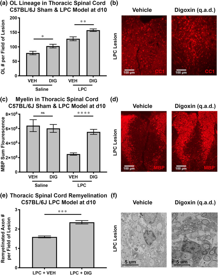
FIGURE 4.

Digoxin promoted robust recovery of myelin in the LPC C57BL/6J mouse model of spinal cord focal demyelination/remyelination. Saline or LPC was injected into the thoracic spinal cord of 10‐week‐old female C57BL/6J mice on day 0. Performed blinded, mice were then treated i.p. with vehicle (VEH) or digoxin (DIG) 0.3 mg/kg qad on days 1–9 and sacrificed on day 10. Immunohistochemical quantitation of the average numbers of OLs (CC1+) was determined for 3 mice/group (3–4 20× images per mouse) (a, b). Immunofluorescent staining of myelin basic protein was carried out with representative images for the treatment groups shown and MBP staining quantitated with results presented as fluorescence intensity (n > 100 values/mouse, n = 3 mice/group) (c, d). Remyelination was assessed by scanning EM of thoracic spinal cord at d10 post‐LPC injection and the mean number of remyelinated axons were determined in n > 12 sections (10,000×, grid square area = 86.61 mm2) of lesion (external and internal) evaluated for each mouse in n = 4 mice/group (e, f). (*p < .05, **p < .01, ***p < .001, ****p < .0001, ns, not significant). Extended data for Figure 4 is labeled as Figure S2.
