
Figure 2.
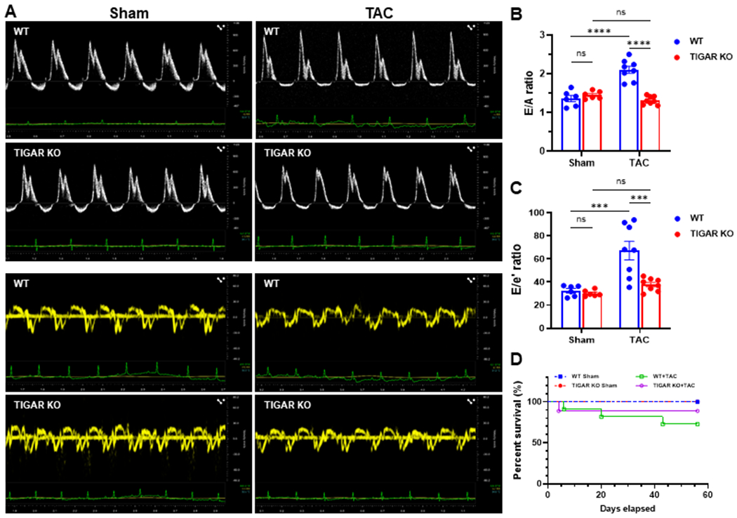
Knockout of TIGAR preserves diastolic function. A, The representative pulsed-wave Doppler and tissue Doppler images from an apical 4-chamber view of WT and TIGAR KO mice subjected to either sham or TAC procedure for eight weeks. B, The ratio of the peak velocity of early (E) to late (A) filling of mitral inflow (E/A) in the indicated groups (n=6–8). C, The ratio of E to the tissue motion velocity in early diastole (e’) was calculated in the indicated groups (n=6–8). D, Kaplan-Meier survival curve. There was no significant difference in survival between the WT and TIGAR KO mice after TAC. ***p<0.001, ****p<0.0001.