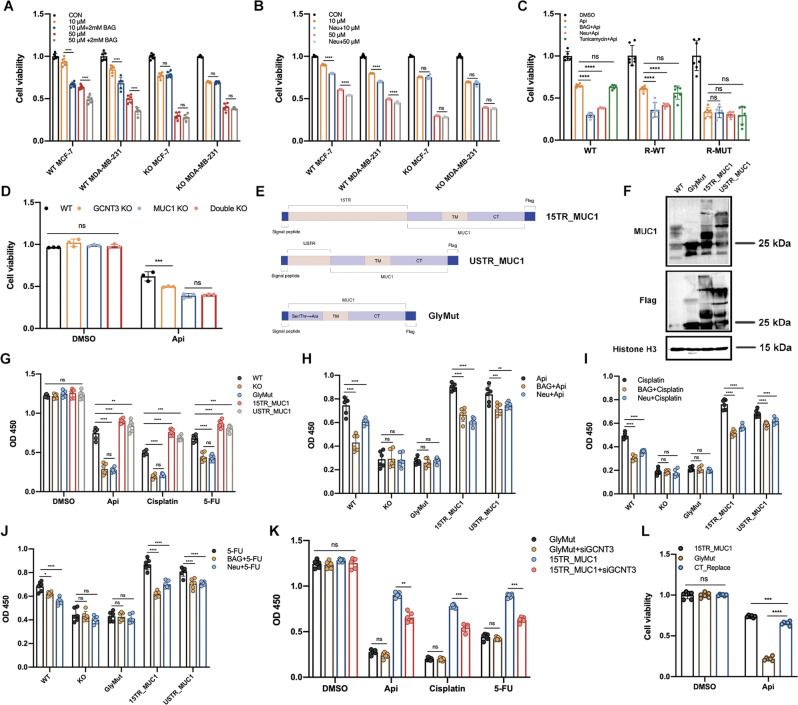
Fig. 4. MUC1 glycosylation inhibition re-sensitized MCF-7 cells to drug cytotoxicity.
A Cell viability of WT/KO cells pretreated with 2 mM BAG post apigenin treatment for 48 h. Both MCF-7 and MDA-MB-231 were detected. B Cell viability of WT/KO cells pretreated with Neu post apigenin treatment for 48 h. Both MCF-7 and MDA-MB-231 were detected. C Cell viability of WT, R-WT, and R-MUT cell lines pretreated with BAG, Neu or tunicamycin after 50 μM apigenin treatment for 48 h. The concentration of BAG was 2 mM and tunicamycin was 0.5 ug/ml. D Cell viability of four cell lines (WT, GCNT3 KO, MUC1 KO, and Double KO) treated with 50 μM apigenin for 48 h. E The diagram of plasmids construction. F Western blot verification of MUC1 O-glycosylation in WT, GlyMut, 15TR_MUC1, and USTR_MUC1 cell lines. G OD value of five cell lines after drug treatments. H OD value of five cell lines pretreated with BAG or Neu post apigenin treatment for 48 h. I OD value of five cell lines pretreated with BAG or Neu post cisplatin treatment for 48 h. J OD value of five cell lines pretreated with BAG or Neu post 5-FU treatment for 48 h. K Cell viability of GlyMut and 15TR_MUC1 with/without siGCNT3 treated with indicated drugs. L Cell viability of 15TR_MUC1, GlyMut, and CT_Replace cells treated with DMSO or 50 μM apigenin for 48 h. Each group was analyzed in triplicate. *P < 0.05; **P < 0.01; ***P < 0.001 for comparisons.

