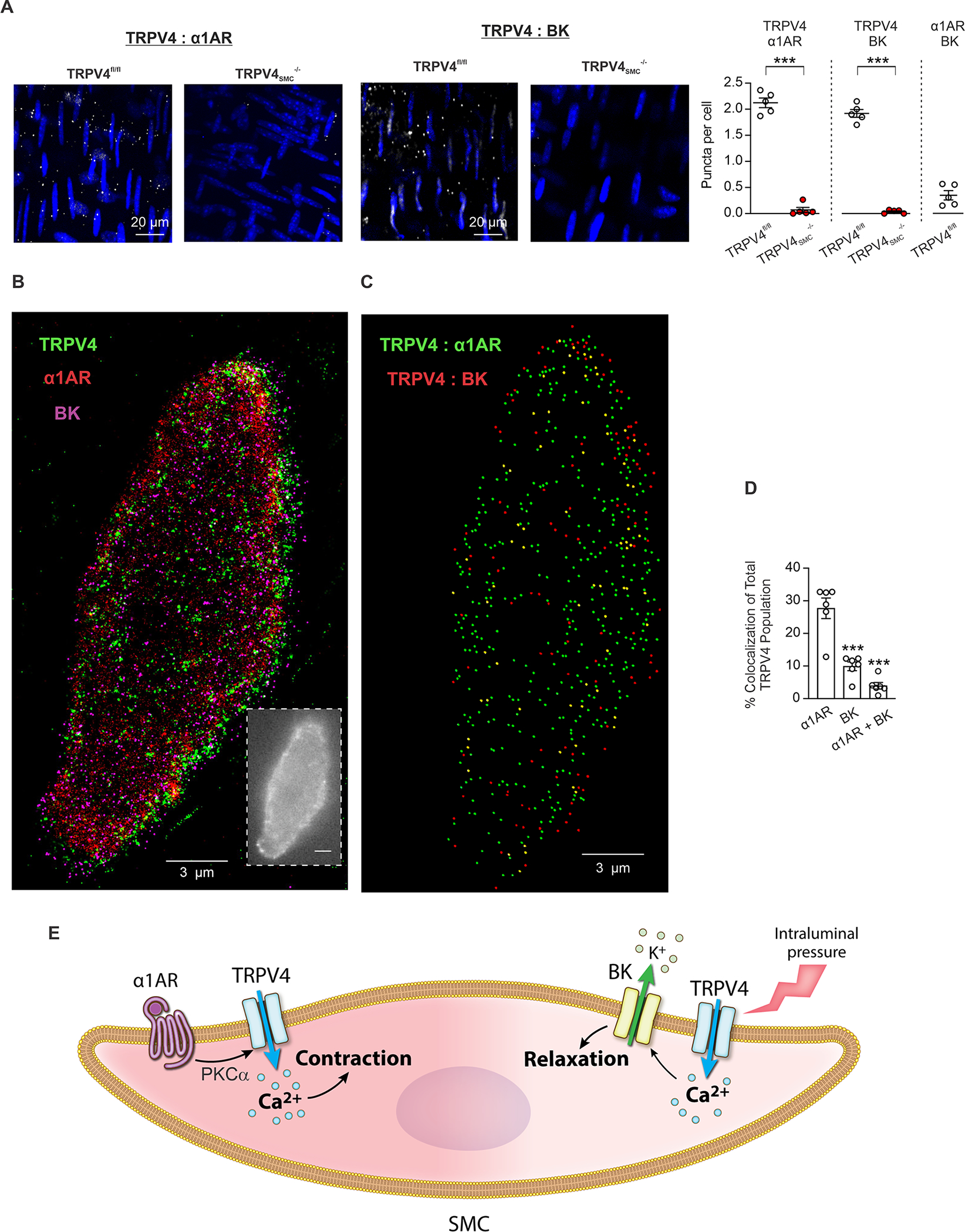
Fig. 4. Discrete α1AR:TRPV4SMC and TRPV4SMC:BK channel signaling nanodomains in SMCs.

(A) Left, representative in situ proximity ligation assay (PLA) images showing SMC nuclei (blue) and α1AR:TRPV4SMC co-localization (white puncta) in en face preparations of MAs from TRPV4fl/fl (left) and TRPV4SMC−/− (right) mice. Middle, representative PLA images of SMC nuclei (blue) and TRPV4SMC:BK co-localization (white puncta) in en face preparations of MAs from TRPV4fl/fl (left) and TRPV4SMC−/− (right) mice. Right, quantification of α1AR:TRPV4SMC (left) and TRPV4SMC:BK co-localization (right) in MAs from TRPV4fl/fl and TRPV4SMC−/− mice (***P < 0.001 vs. TRPV4fl/fl; unpaired t-test). (B) Superresolution localization maps for TRPV4, α1AR and BK channels in native SMCs from control mice. The inset shows a widefield snapshot of one SMC. Scale bars, 3 μm. (C) Co-localization analysis (Imaris) results for localization of TRPV4 channels with α1ARs (green dots), BK channels (red dots), or both (yellow dots). (D) Summary of co-localization analysis showing the percentage of total TRPV4 channels coupling with α1ARs, BK channels, or both in SMCs from control mice (n = 6; ***P < 0.001 vs. α1AR-paired; one-way ANOVA). (E) Schematic diagram showing the divergent roles of TRPV4SMC channels in regulating arterial diameter. Pressure-induced TPRV4SMC:BK channel signaling causes vasodilation. In contrast, α1AR:TPRV4SMC channel signaling elicits vasoconstriction.
