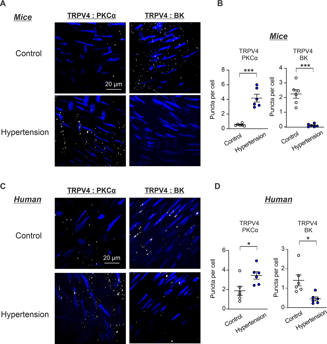
Fig. 7. PKCα:TRPV4SMC co-localization is increased and TRPV4SMC:BK co-localization is reduced in hypertension.

(A) Representative in situ PLA images showing SMC nuclei (blue), TRPV4SMC:PKCα and TRPV4SMC:BK co-localization (white puncta) in en face preparations of MAs from Ang II-induced hypertensive mice and non-hypertensive control mice. (B) Quantification of TRPV4SMC:PKCα (Control, n = 6; Hypertension, n = 6) and TRPV4SMC:BK co-localization (Control, n = 6; Hypertension, n = 6) co-localization in MAs from Ang II-induced hypertensive mice and control mice (***P < 0.001 vs. Control; unpaired t-test). (C) Representative in situ PLA images showing SMC nuclei (blue), TRPV4SMC:PKCα and TRPV4SMC:BK co-localization (white puncta) in en face preparations of paraspinal muscle arteries from non-hypertensive (control) and hypertensive individuals. (D) Quantification of TRPV4SMC:PKCα (Control, n = 6; Hypertension, n = 6) and TRPV4SMC:BK (Control, n = 6; Hypertension, n = 6) co-localization in paraspinal muscle arteries from non-hypertensive (control) and hypertensive individuals (*P < 0.05 vs. Control; unpaired t-test).
