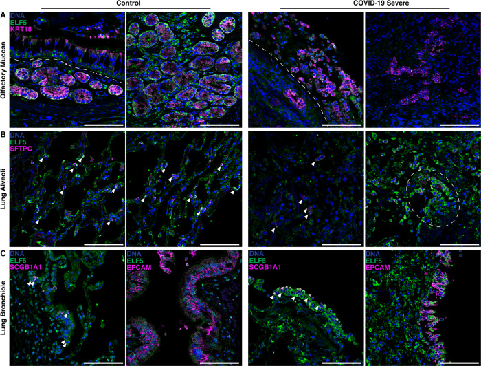
Fig. 5. ELF5 expression by epithelial cells of the olfactory mucosa and lung.
Immunofluorescent staining of ELF5 in control and COVID-19 patients in the A olfactory mucosa, B lung alveoli, and C lung bronchiole. A Dashed lines separate the olfactory epithelium and the lamina propria. B Arrowheads highlight AT2 cells expressing ELF5; dashed outline highlights clusters of AT cells expressing ELF5. C left: epithelial cells expressing ELF5; right: arrowheads highlight airway epithelial cells expressing ELF5. Marker genes for sustentacular and Bowman gland cells (A KRT18), alveoli type II cells (B SFTPC), pan-epithelial cells (C EPCAM), and secretory cells (C SCGB1A1) are shown in purple. Validation staining for each tissue: control (n = 2); COVID-19 (n = 2). Scale bar = 100 μm.

