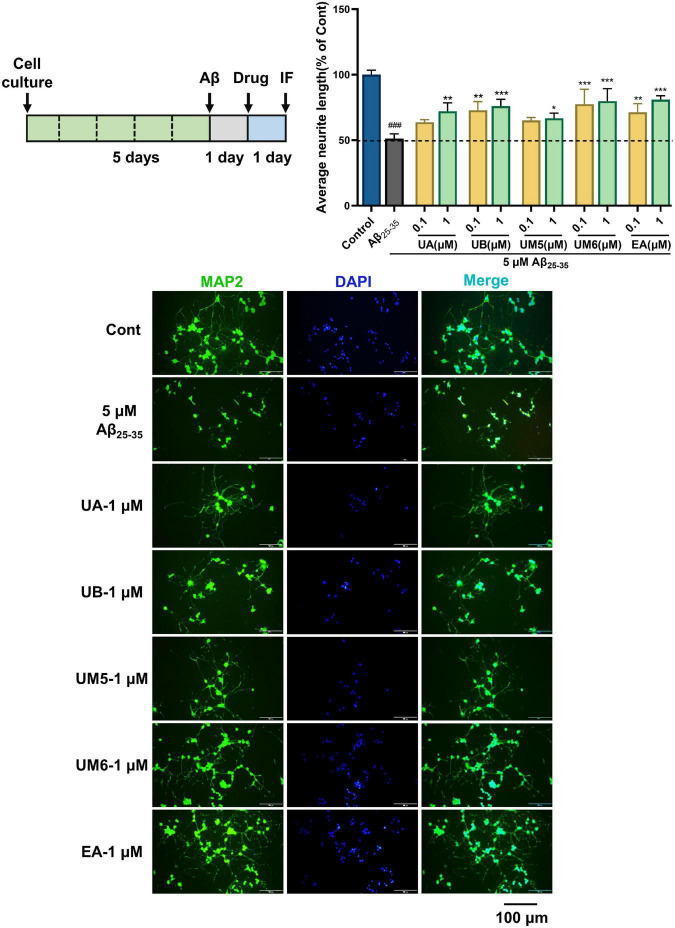
FIGURE 4.
Effects on Aβ25–35-induced neurite atrophy in PC12. The PC12 cells were differentiated for 5 days, and then the cells were treated with (Aβ25–35) or without (Cont) 5 μM Aβ25–35 for 24 h. Each compound at a concentration of 0.1 or 1 μM containing NGF (100 ng/mL), or Aβ25–35 was added to the neurons and cultured for another 24 h after removing the Aβ-containing medium. Then the cells were fixed and immunostained with MAP2 and DAPI. The lengths of the MAP2 positive neurites were measured. The values are the means ± S.D. of the data. n = 3. ### p < 0.001 vs. Control, *p < 0.05 vs. Aβ25–35, **p < 0.01, ***p < 0.001. Scale bar = 100 μm.

