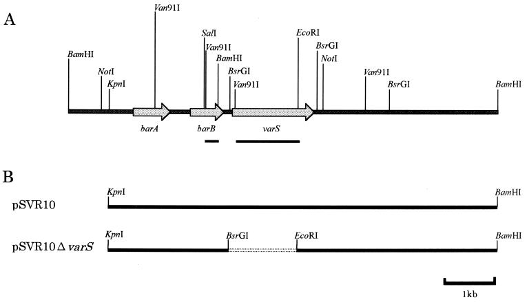
FIG. 2.
(A) Restriction map of an 8.2-kb BamHI fragment containing varS and the upstream and downstream regions. ORFs corresponding to barA, barB, and varS are indicated by shaded arrows. Probes (1.19-kb Van91I-EcoRI fragment for varS and 262-bp SalI-BamHI fragment for barB) used for Northern blot hybridization are indicated by filled boxes below the arrows. (B) Schematic representation of the inserts in pSVR10 and pSVR10ΔvarS used for the in vivo functional analysis of varS. Inserts were first constructed in pUC18, recovered as HindIII-XbaI fragments by use of the corresponding flanking restriction sites of pUC18, and then ligated into HindIII-XbaI-digested pIJ486. Broken lines indicate the deletion of varS.

