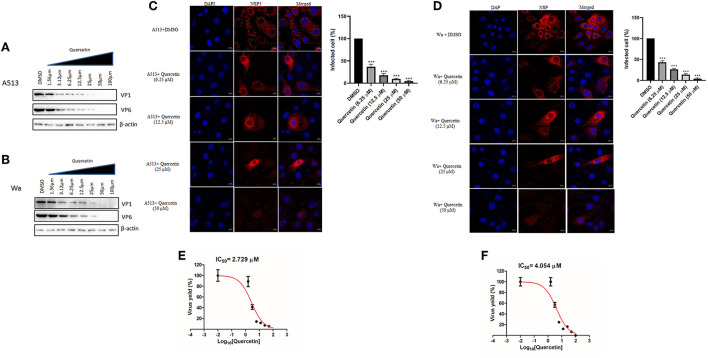
Figure 2.
Quercetin exhibits anti-rotaviral activity independent of viral strain: (A) Cell lysates were prepared to perform Western blot by infecting MA104 cells with RV-A513 strain followed by treatment with DMSO or increasing concentration of quercetin (1.56, 3.125, 6.25, 12.5, 25, 50, and 100 μM) to assess viral protein expression. The expression of β-actin is checked as internal loading control. (B) Similarly, RV-Wa strain-infected MA104 cells are treated with DMSO or increasing doses of quercetin (1.56, 3.125, 6.25, 12.5, 25, 50, and 100 μM) to check viral proteins along with β-actin as loading control. (C,D) MA104 cells were infected with (C) RV-A5-13 and (D) RV-Wa (MOI 3) in the presence of DMSO and quercetin (6.25, 12.5, 25, and 50 μM) for 6 hpi. Cells were fixed, permeabilized, and stained primarily with anti-NSP5 antibody (raised in rabbit). Next, secondary staining was done with rhodamine-labeled anti-rabbit secondary antibody. DAPI was used for mounting. Finally, cells were visualized with confocal microscope (63× oil immersion); scale bar 10 μm. Viroplasm positive cells are quantified and analyzed by taking into account minimum 100 cells selected randomly from each experimental slides. The data were represented as mean-infected cells (%) ± SD of three experimental replicates. (E,F) IC50 was calculated from the quantification of viral particle produced by plaque assay which was performed from (E) A5-13- and (F) Wa-infected MA104 cells treated with DMSO and graded concentration of quercetin (1.56, 3.125, 6.25, 12.5, 25, 50, and 100 μM), for 24 hpi. Each bar represents mean ± SD of three independent experiments (Unpaired student's t-test, ***p < 0.001).

