. 2022 Jun 24;82(16):2838–2847. doi: 10.1158/0008-5472.CAN-22-0554

Figure 1.

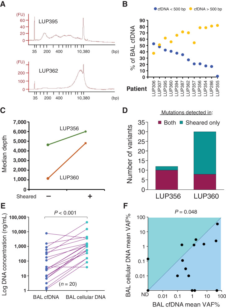
Figure 1. Characteristics of BAL DNA from lung cancer. A, cfDNA fragment size distribution for two representative BAL cfDNA samples. FU, fluorescence units. B, DNA fragment size for BAL cfDNA stratified by a 500-bp threshold (orange > 500 bp, blue < 500 bp) for n = 12 patients (x-axis). C, Median sequencing depth obtained from BAL cfDNA samples with and without shearing. D, Number of mutations detected in both sheared and unsheared samples or in sheared samples alone for the two patients from (C). A total of 42 mutations were detected in sheared samples versus 18 in unsheared (P < 0.001). E, BAL DNA concentrations in 20 patients with lung cancer for BAL cfDNA and BAL cellular DNA. F, Comparison of mean VAF% for BAL cfDNA and BAL cellular DNA for 15 patients. ND, not detected. P is displayed above the plot.

Characteristics of BAL DNA from lung cancer. A, cfDNA fragment size distribution for two representative BAL cfDNA samples. FU, fluorescence units. B, DNA fragment size for BAL cfDNA stratified by a 500-bp threshold (orange, >500 bp; blue, <500 bp) for n = 12 patients (x-axis). C, Median sequencing depth obtained from BAL cfDNA samples with and without shearing. D, Number of mutations detected in both sheared and unsheared samples or in sheared samples alone for the two patients from C. A total of 42 mutations were detected in sheared samples versus 18 in unsheared (P < 0.001). E, BAL DNA concentrations in 20 patients with lung cancer for BAL cfDNA and BAL cellular DNA. F, Comparison of mean VAF% for BAL cfDNA and BAL cellular DNA for 15 patients. ND, not detected. P is displayed above the plot.