
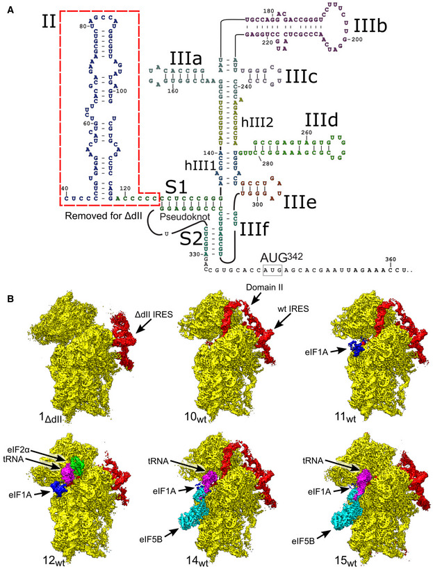
Figure 1. Overview of HCV IRES‐mediated initiation complexes.
-
ASecondary structure of the HCV IRES, annotated to show individual elements.
-
BSegmented maps of the indicated ribosomal complexes assembled on the wt or ΔdII HCV IRES, showing the 40S subunit (yellow), IRES (red), eIF1A (blue), Met‐tRNAi Met (magenta), and initiation factors eIF2 (green) or eIF5B (cyan).