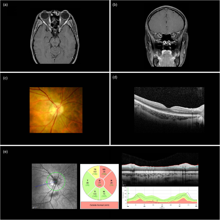
Figure 1.
Ocular findings in case 1. (a) Axial T1 MRI of brain and orbits, with fat suppression sequences and post gadolinium contrast, demonstrating enhancement of the retrobulbar right optic nerve near the optic foramen. (b) Coronal T1 MRI of brain and orbits, with fat suppression sequences and post gadolinium contrast, demonstrating enhancement of the retrobulbar right optic nerve. (c) Fundus photo of right optic disc characterized by peripapillary atrophy and mildly blurred margins. (d) Macular OCT scan of the right eye. (e) Retinal nerve fiber layer (RNFL) thickness measurements of right eye using OCT.

