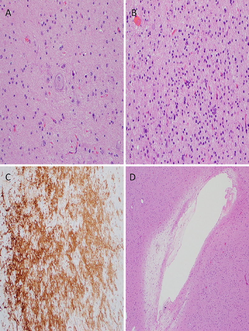
FIG. 2.
Histological sections of the tumor with representative features. A: Dystrophic neuron (H&E stain, original magnification ×200). B: Hypercellularity (H&E stain, original magnification ×200). C: CD34+ cell populations (immunohistochemistry stain, original magnification ×200). D: Microcavitations (H&E stain, original magnification ×100).

