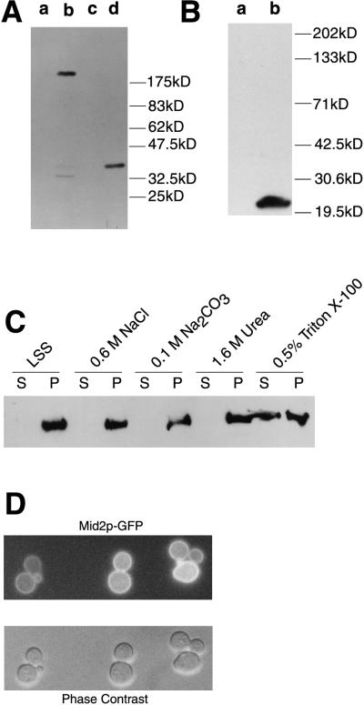
FIG. 2.
Cell biology of Mid2p. (A) Immunoblot analysis of cell extracts from TK82 (vector only) (lane a), TK84 (MID2-HA) (lane b), pmt1Δ pmt2Δ (vector only) (lane c), and pmt1Δ pmt2Δ (MID2-HA). (B) Immunoblot analysis of cell extracts from TK82 (vector only) (lane a) and TK85 (ΔS/T-Mid2p-HA) (lane b). (C) Immunoblot analysis of cell fractions from TK84 to demonstrate membrane association of Mid2p. LSS, low-speed-spin pellet fraction. (D) In cells expressing pRS426-MID2-GFP (TK98), Mid2p-GFP is localized to the cell periphery.

