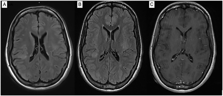
Figure 1.
Axial T2 FLAIR showed cortical and subcortical hyperintensities within the brain parenchyma involving the left middle frontal gyrus, right superior frontal gyrus, right parietal cortex, left insula and right occipital lobe, and multiple subependymal nodules (A, B). Axial T1 post-contrast (C) showed enhancing subependymal nodule at the right foramen of Monro and smaller ring enhancing subependymal nodule along the posterior periventricular right white matter.

