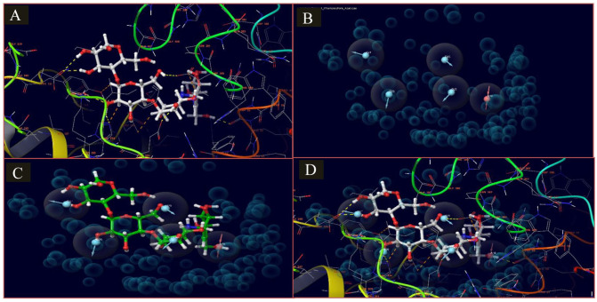
Figure 3.
E-pharmacophore model of the IFD docked complex of acarbose and apo crystal structure of HPA (6OCN_A). (A) This shows the 3D representation of the docked complex and the interaction of acarbose atoms in the active site of HPA (6OCN_A). (B) The generated e-pharmacophore model is based on the stabilized and energy-minimized interaction in the complex. (C) Pharmacophore model on the interacting atoms of acarbose. (D) Pharmacophore model in the protein complex of acarbose and HPA (6OCN_A).
HPA indicates human pancreatic amylase; IFD, induced-fit docking.

