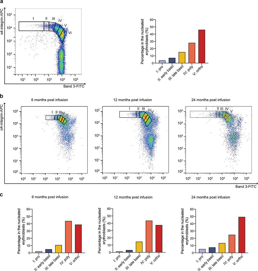
Extended Data Fig. 3 |. Erythropoietic maturation in bone marrow (Patient 2).
Terminal erythroid differentiation begins with proerythroblasts differentiating into basophilic, then polychromatic, then orthochromatic erythroblasts that enucleate to become reticulocytes. Each distinct stage of terminal human erythroid differentiation can be distinguished using a combination of cell surface markers for glycophorine A (GPA), band 3 and α4 integrin. a, Representative flow cytometry plot of band 3 vs α4-integrin of GPA+ cells in normal erythopoesis: proerythrobalsts (I), early basophilic (II), late basophilic (III), polychromatic (IV), and orthochromatic erythroblasts (V) and reticulocytes (VI). The box plot represents the quantitation of the proportion of cells at each distinct stage of maturation after normalization based on total nucleated erythroid cells (I-V) as 100% as described in ref29. The left panel is adapted from ref29.
b, Bone marrow erythroblasts from Patient 2 were analyzed by flow cytometry at 6, 12 and 24 months post infusion stained with GPA, α4-integrin, and band 3. Plot of band 3 vs α4-integrin of GPA+ cells represents the quantitation of distinct stages of maturation of erythroblasts as described in a. c, Quantitation of the proportion of cells at each distinct stage of maturation after normalization to total nucleated erytroid cells (I-V).

