
Figure 6.
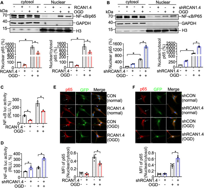
RCAN1.4 suppresses OGD‐induced NF‐κB/p65 nuclear translocation in primary astrocytes. (A and B) Cultured primary astrocytes infected with Lenti‐RCAN1.4‐GFP or Lenti‐shRCAN1.4‐GFP for 5 days were treated with or without OGD for 6 h. Nuclear proteins were extracted from cells infected with Lenti‐RCAN1.4‐GFP or Lenti‐shRCAN1.4‐GFP. NF‐κB/p65 proteins were detected using an anti‐p65 antibody. GAPDH and H3 were used as a loading control for the cytosolic and nuclear fractions, respectively. (C and D) The RCAN1.4 expression construct or silencing plasmid was cotransfected with pNF‐κBLuc into HEK293 cells. A dual luciferase assay was performed at 12 h after OGD treatment. (E and F) Cultured primary astrocytes infected with Lenti‐RCAN1.4‐GFP or Lenti‐shRCAN1.4‐GFP for 5 days were treated with or without OGD for 6 h. The cells were fixed and permeated, then stained with anti‐p65 (Alexa Fluor 594, red) and DAPI for the nucleus (blue). The image was captured with Leica confocal microscopy (LSM880). Scale bar 10 μm. Values represent means ± SEM (n = 3), *p < 0.05, as calculated by one‐way ANOVA with Bonferroni's multiple comparison post hoc test. MFI, mean fluorescence intensity; OGD, oxygen–glucose deprivation; RCAN1.4, the isoform 4 of regulator of calcineurin 1.