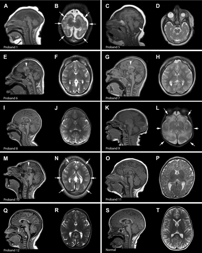
Figure 3.

Brain imaging in ANKLE2‐related microcephaly. Select brain MR images of individuals with ANKLE2 variants are shown. (A and B) (proband 1), diffuse and severe undersulcation of the cortical gyral pattern with thin‐normal cortical thickness (arrows), increased extra‐axial space (asterisks), severe white matter involvement, dysplastic ventricles with complete agenesis of the corpus callosum with open communication between the trigones and medial extra‐axial space; (C and D) (proband 5), diffuse simplification and undersulcation of the cortical gyral pattern, partial agenesis of the corpus callosum (arrowhead), cerebellar vermis hypoplasia (arrow) (limited images available); (E and F) (proband 6), diffuse simplification of the cortical gyral pattern with severely foreshortened frontal lobes, very short and thick corpus callosum with partial agenesis (arrowhead), relatively preserved brainstem and cerebellum; (G and H) (proband 7), similar appearance as the sibling (proband 6) with diffuse simplification of the cortical gyral pattern, partial agenesis of the corpus callosum (arrowhead), and relatively preserved brainstem and cerebellum; (I and J) (proband 8), mild simplification of the cortical gyral pattern with mild foreshortening of the frontal lobes and mildly thin corpus callosum; (K and L) (proband 9), diffuse and severe simplification of the cortical gyral pattern (arrows), diffuse white matter abnormalities, mildly increased extra‐axial spaces; (M and N) (proband 10), diffuse severe simplification of the cortical gyral pattern (arrows), severe foreshortening of the frontal lobes, partial agenesis of the corpus callosum (arrowhead), relatively preserved brainstem and cerebellum; (O and P) (proband 11), mildly thin corpus callosum, with mild simplification of the cortical gyral pattern; (Q and R) (proband 12), diffusely simplified gyral pattern with paucity of the sulci in various areas, mild reduction in supratentorial white matter and a cisterna magna; (S and T) (normal), normal mid‐sagittal and axial images.
